
Four Treasures Reference Database
Record No. 5058
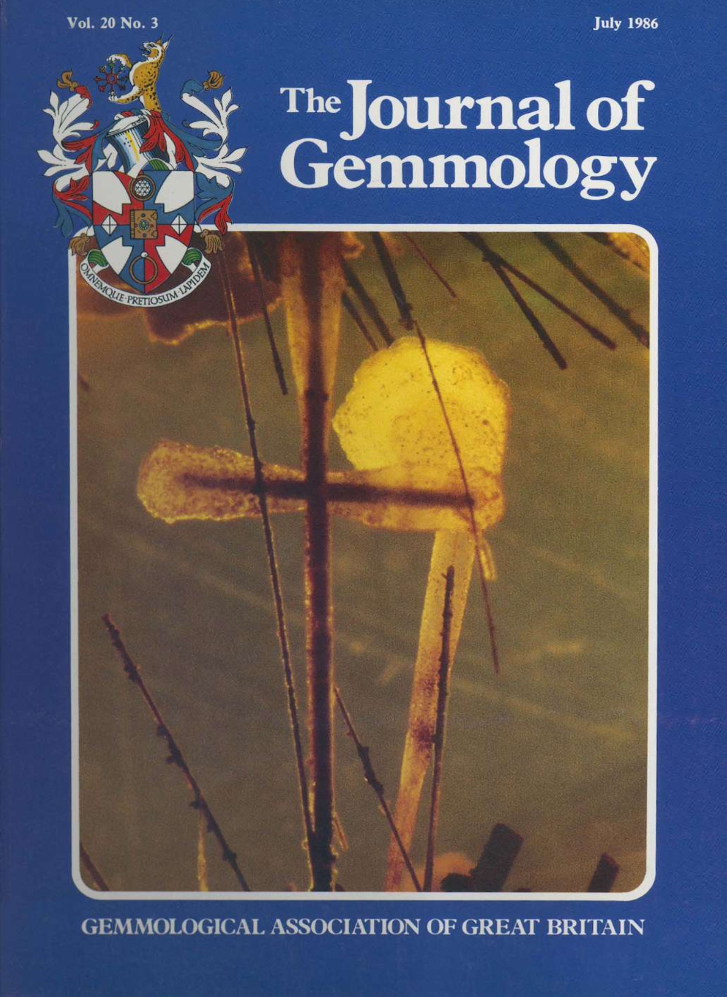
Inclusions in opal
Journal Article
Gübelin, E.J. and Koivula, J.I. (1986) Inclusions in opal. Journal of Gemmology, Vol. 20, No. 3, July, pp. 139–144.
ISBN/ISSN: 221252 | Sinkankas/Born No.: NIS / NIB | RWHL
opal, inclusions, gems
This illustrated survey describes the diversity, appearance and geological significance of inclusions in natural opal, drawing principally on transparent and translucent Mexican material. Published as a preview of the authors’ Photoatlas of Inclusions in Gemstones, it combines an introductory account of opal with 26 photomicrographs showing mineral inclusions, internal structures, fluid cavities and treatment features. Many opals lack visible inclusions or are too opaque for their inclusions to be examined, whereas Mexican fire and water opals provide particularly favourable opportunities for observation. Hornblende needles and crystals, goethite, quartz, chalcedony, cristobalite, kaolinite and fragments of rhyolite matrix occur in varied associations. Pre-existing hornblende and goethite crystals projecting from cavity walls were enclosed during opal deposition, and some retain coatings or alteration products. Successive deposition around earlier crystals produces white opal envelopes and concentric structures resembling tree rings. A broken needle-like inclusion is interpreted as evidence of movement within the viscous silica gel. Chalcedony forms rounded or botryoidal masses, sometimes accompanied by curved fractures attributed to internal stress, while cristobalite occurs as white inclusions or aggregates of minute plate-like crystallites. Quartz crystals may project from kaolinite or the rhyolite matrix and may carry limonite coatings. Hornblende can likewise be coated or replaced by limonite, producing distinctive pseudomorphs. A three-phase inclusion contains liquid, a large gas bubble and numerous small red solids within a rhombohedral cavity interpreted as the former site of a mineral such as calcite or dolomite. Later alteration is illustrated by an iron-rich deposit left in a fracture after invading fluids dried out. Examples from other localities include manganese oxide dendrites in Tanzanian yellow opal, tiny dark grey bravoite octahedra reported in Brazilian white opal, and sand-like matrix particles in Australian material. Finely laminated botryoidal structures in Mexican hyalite produce iridescence, while Australian oolitic opal displays natural circular structures that should not be mistaken for treatment effects. The final examples distinguish these natural features from black subsurface soot clouds in smoke-treated Mexican opal and the pepper-like appearance produced by sugar and sulphuric acid treatment of porous Australian opal. The survey demonstrates how microscopy reveals successive stages of inclusion entrapment, opal deposition and alteration, while also helping recognise artificial darkening.