The phenomenon of pleochroism in doubly refractive (DR) gemstones is described, as it relates to overall color appearance in the context of faceted gems. With strongly pleochroic gems, pleochroism can be seen in faceted gems, even if the table facet is oriented perpendicular to an optic axis. This is because facets change the direction of light as it moves through the gem.
Introduction
In describing colored gemstones, the seven major factors influencing color are: hue position, lightness (tone), saturation (intensity), color zoning, color-change (color inconstancy), dispersion and pleochroism. This article deals with the last of these – pleochroism.
The term pleochroism (from the Greek “pleio,” many, and “chros,” color) describes a variation of color with direction in doubly refractive gems (Figure 1). Virtually all gemological texts note this effect and describe it in detail. Yet many of these descriptions deal only with the identification aspects of this property. When lapidary aspects are discussed, the focus is on orientation, generally omitting mention of exactly how pleochroism affects the appearance of faceted stones. The purpose of this article is to provide a detailed description of pleochroism as it relates to the appearance of faceted gems.
 Figure 1. Pleochroism in a 15-ct tanzanite as seen with the naked eye through the crown, side and end.
Photos: Wimon Manorotkul
Introduction to Optical Crystallography
For gemological purposes, crystals can be divided into three optical groups based on their lattice symmetry: isometric, dimetric and trimetric, meaning crystals that are built of equidimensional lattice points (isometric), those that are stretched or compressed in one dimension but equidimensional in the other two (dimetric) and those where each of the three dimensions is different (trimetric).
Imagine a perfect sphere. If you draw a line from the center to the surface in any direction, all possible lines will have the same length. This sphere represents how light behaves in isometric crystals (iso = one; metric = length).
Now if we stretched or squashed that perfect sphere in a single direction, it would become dimetric. A perfect circle when viewed in one direction, but oval when viewed at right angles to the direction of distortion. In other words, two of the three dimensions are equidistant, but the third is either stretched (uniaxial +) or compressed (uniaxial –).
Finally, if we distort our perfect sphere not just in one dimension, but all three, we end up with a trimetric crystal, where each of the three dimensions differs from the other (Figure 2).
The optical properties of crystals are summarized in Table 1.
| Structure | Structure Type | Optic character | Pleochroism | Examples |
|---|---|---|---|---|
| Amorphous | No order No axes |
Singly refractive | None | Amber Glass |
| Isometric | One crystallographic axis length (cubic) a1 = a2 = a3 |
Singly refractive | None | Diamond Spinel |
| Dimetric | Two crystallographic axis lengths a1 = a2 ≠ c (tetragonal) a1 = a2 = a3 ≠ c (hexagonal) |
Doubly refractive Uniaxial Two RI’s (nω, nε), one optic axis |
May be dichroic | Corundum Tourmaline Zircon |
| Trimetric | Three crystallographic axis lengths a ≠ b ≠ c (orthorhombic, monoclinic, triclinic) |
Doubly refractive Biaxial Three RI’s (nα, nβ, nγ), two optic axes |
May be trichroic | Andalusite Iolite Topaz Zoisite |
Within singly refractive (isotropic) gem materials, the spacing of lattice points is essentially the same in all three mutually perpendicular dimensions (top to bottom, side to side, back to front). As a result, these materials transmit light in a manner that is independent of crystal orientation. Light moves through them at a single speed and is permitted to vibrate in any direction perpendicular to the direction of travel.
This is not the case, however, in doubly refractive (anisotropic) gems. All doubly refractive gems are anisotropic crystals, and the arrangement of their lattice points varies with direction. Uniaxial materials possess one unique direction (the c axis) along which the spacing of lattice points differs from that of the other crystal axes (the a axes); with biaxial gems, the spacing differs along each of the three crystal axes (a, b and c axes). The result of this differing arrangement of lattice points in doubly refractive gems is that light is split into two separate rays (with the exception of rays travelling parallel to an optic axis) that each travel at different speeds and thus possess different refractive indices (RI's). For more on the behavior of light in crystals, please see Box A.
Uniaxial gems possess two rays—the ordinary (o) and extraordinary rays (e)—and two RI values (omega and epsilon, respectively). The o-ray always vibrates perpendicular to the c axis and the e-ray vibrates parallel to the c axis. The vibrations of each ray are mutually perpendicular, and they vibrate roughly perpendicular to the direction of travel.
In biaxial gems, there are three different rays and three different RI's (alpha, beta and gamma), although only two are ever found in any one direction. The vibrations of alpha, beta and gamma are parallel to the three optical directions, X, Y, and Z, respectively. Although the three crystallographic axes of biaxial minerals are not always mutually perpendicular, the three optical directions – X, Y and Z – are always 90° to one another.
Uniaxial gems contain one optic axis (direction of single refraction), which is parallel to the c axis; biaxial gems contain two optic axes (two directions of single refraction), which lie in the X-Z plane. The major vibration directions of both uniaxial and biaxial gems are shown in Figure 2.
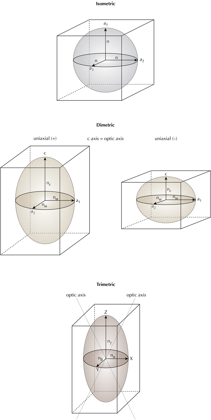 Figure 2. Vibration directions of light waves in crystals. Isometric crystals do not split light and thus display no pleochroism. Uniaxial crystals possess two vibration directions, allowing the possibility of two pleochroic colors (dichroism), while biaxial crystals have three vibration directions and thus the potential to display three pleochroic colors (trichroic). In both uniaxial and biaxial crystals, the major optical directions are always mutually perpendicular, which is not necessarily the case for the crystallographic axes in crystals (only cubic, tetragonal and orthorhombic crystals have exclusively mutually perpendicular crystallographic axes). Illustration by Richard W. Hughes.
Because each polarized ray in a doubly refractive gem is vibrating across a slightly different arrangement of lattice points, each ray is absorbed differently. In many cases, the difference in absorption between one ray and another is too slight to detect with the eye, or even with a dichroscope. However, where the differences are large enough, this will result in a variation in color with direction. Such variations are known as pleochroism, and are most distinct in deeply colored specimens of the same gem species.
Uniaxial gems possess two different vibrations – omega (ω) and epsilon (ε) – also known as the ordinary and extraordinary rays – and may therefore exhibit two different colors (termed 'dichroism'). Biaxial gems possess three different vibrations – alpha (α), beta (β) and gamma (γ) – and so may show three different colors (termed 'trichroism'), but only two in any single direction. Blue sapphire and tourmaline are examples of strongly dichroic uniaxial gems, while iolite provides an example of a strongly trichroic biaxial stone. Not all uniaxial gems show visible dichroism, however, and not all biaxial gems display visible trichroism. Some uniaxial gems may show only one color, while biaxial gems may show only one or two colors.
When viewed in transmitted light with a dichroscope, pleochroism in uniaxial stones varies in the following ways, as shown in Figure 3:
- Parallel to the c-axis, only the ordinary ray is seen and thus there is no pleochroism
- At right angles to the c axis, both the ordinary and extraordinary rays are seen; this position shows the strongest dichroism
- At angles oblique to the c axis, the ordinary ray is seen; the second color (usually referred to as ε') diverges from that of the o-ray as one moves away from the c axis
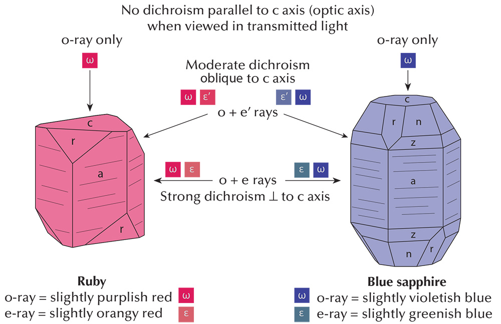 Figure 3. Pleochroism in uniaxial crystals as viewed with the dichroscope, using the example of ruby and sapphire. Illustration: Richard W. Hughes
With biaxial stones, the situation is similar, but slightly more complex. Light traveling along the Z direction will vibrate parallel to X and Y, and so the two colors seen with a dichroscope will be those of alpha (X) and beta (Y). Light traveling along the X direction will vibrate across Y and Z, and so the two colors seen will be those of beta (Y) and gamma (Z). When light travels parallel to Y, it vibrates along X and Z; thus the colors seen are those of alpha (X) and gamma (Z). Should light travel parallel to either of the two optic axes in a biaxial stone, only one color will be seen, that of beta. This is illustrated in Figure 4.
.jpg) Figure 4. Pleochroism in biaxial crystals, as viewed with a dichroscope in each of the three major optical directions. When viewed down either of the two optic axes (right), only the beta color will be seen. Note: In biaxial crystals, the two optic axes always lie in the X-Z plane. The smaller (acute) of the two angles between the optic axes in a biaxial stone is called the 2V angle. When the smaller angle is bisected by the Z direction, the crystal is termed 2Vz and is positive in sign. If the smaller of the two angles is bisected by the X direction (2Vx), the crystal is negative in sign. The example above at right is 2Vz, and thus positive in sign. Should the 2V angle be 90°, the optic sign would be neutral (±). Illustration: Richard W. Hughes. Click on the illustration for a larger view.
Figure 4. Pleochroism in biaxial crystals, as viewed with a dichroscope in each of the three major optical directions. When viewed down either of the two optic axes (right), only the beta color will be seen. Note: In biaxial crystals, the two optic axes always lie in the X-Z plane. The smaller (acute) of the two angles between the optic axes in a biaxial stone is called the 2V angle. When the smaller angle is bisected by the Z direction, the crystal is termed 2Vz and is positive in sign. If the smaller of the two angles is bisected by the X direction (2Vx), the crystal is negative in sign. The example above at right is 2Vz, and thus positive in sign. Should the 2V angle be 90°, the optic sign would be neutral (±). Illustration: Richard W. Hughes. Click on the illustration for a larger view.
Pleochroism in Faceted Uniaxial Gems
RI variation (see Hurlbut, 1984) is a directly measurable manifestation of the more subjective differences we see with pleochroism. In the sections that follow, I have used RI variation to model pleochroism, based on the assumption that the two are identical in terms of their variation. If one assigns an RI value to each ray, it is no different than assigning a color value to each ray; thus it follows that as the RI varies, the color will also vary.
Let's examine how this applies to the appearance of faceted stones. Figure 5 shows a uniaxial gem cut with the c axis perpendicular to the table facet (since the crown plays little part in the pleochroism of a faceted stone, it will be eliminated from this discussion for simplicity's sake). Suppose the stone is a perfect square cut with only four pavilion facets (marked N, S, E, and W – standing for North, South, East and West), again in the interest of simplicity. The aaxes are contained in the horizontal plane.
For light entering the stone parallel to the c axis, only the o-ray would be obtained. However, upon striking facet W the direction is changed. It then travels in the plane of the a axes and so an equal mixture of the o- and e-rays is obtained. Upon striking facet E the direction is again changed, causing the ray to exit the stone parallel to c. Once more this produces the o-ray only for this portion of its journey. Should the ray strike facet N or S instead, this still would not alter the above combinations.
On first examination it would seem that uniaxial stones cut with the c axis perpendicular to the girdle plane would show one uniform color across their face. However this is not the case in faceted gems because the facet arrangement causes light to change direction during its journey through the gem. In uniaxial gems where the o-ray is more intensely colored than the e-ray, the color near the girdle will appear slightly less intense than that near the culet, because the percentage of e-ray mixing into the color increases as light approaches the girdle.
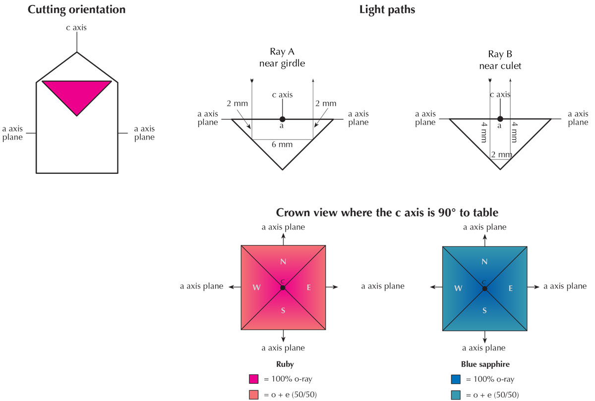 Figure 5. Pleochroism as seen through the crown in ruby and sapphire cut with the table at 90° to the c axis. Note that color variations in figures 5, 6, 8 and 12 are approximate only and designed to show general concepts. Refractive indices and pleochroism vary in logarithmic fashion, while the illustrations were generated showing linear variations. See Hurlbut (1984) for more details. Illustration: Richard W. Hughes
Uniaxial with C-Axis 90° to the Table
Let us examine Figure 5. The girdle diameter of this gem is 10 mm, the pavilion angle is 45°, and the c-axis is cut perpendicular to the table plane.
- Ray A enters near the girdle, travelling 4 mm parallel to the c axis (o-ray only) and 6 mm parallel to the a axis (3 mm of o-ray + 3 mm of e-ray). Thus the color of Ray A consists of 70% o-ray and 30% e-ray.
- Ray B strikes the pavilion much closer to the culet. In terms of path length, Ray A and Ray B are identical. Each ray travels the same 10 mm distance through the gem. Ray B's light path, however, consists of 8 mm parallel to the c axis (8 mm of o-ray only) and just 2 mm parallel to the a axis (1 mm of o-ray + 1 mm of e-ray). Thus the color of Ray B is 90% o-ray and only 10% e-ray.
The conclusion is that when a uniaxial gem is cut with the c axis perpendicular to the table facet, there can still be visible pleochroism. The o-ray color is stronger at the culet, gradually diminishing toward the girdle. To the best of the author's knowledge, this fact has not been described in the gemological literature.
In uniaxial gems such as blue zircon, ruby, blue sapphire and tourmaline, where the e-ray is less saturated in color than the o-ray, the color near the girdle will appear slightly less intense than that near the culet, because the percentage of e-ray mixing into the color increases as light approaches the girdle. With gems such as blue benitoite, where the e-ray is more intensely colored than the o-ray, just the reverse would hold true. The color would be lightest at the culet, darkening toward the girdle. Since making this discovery the author has found that these differences are subtle, at best, and in many cases undetectable. In large, strongly pleochroic stones, however, the effect can be striking.
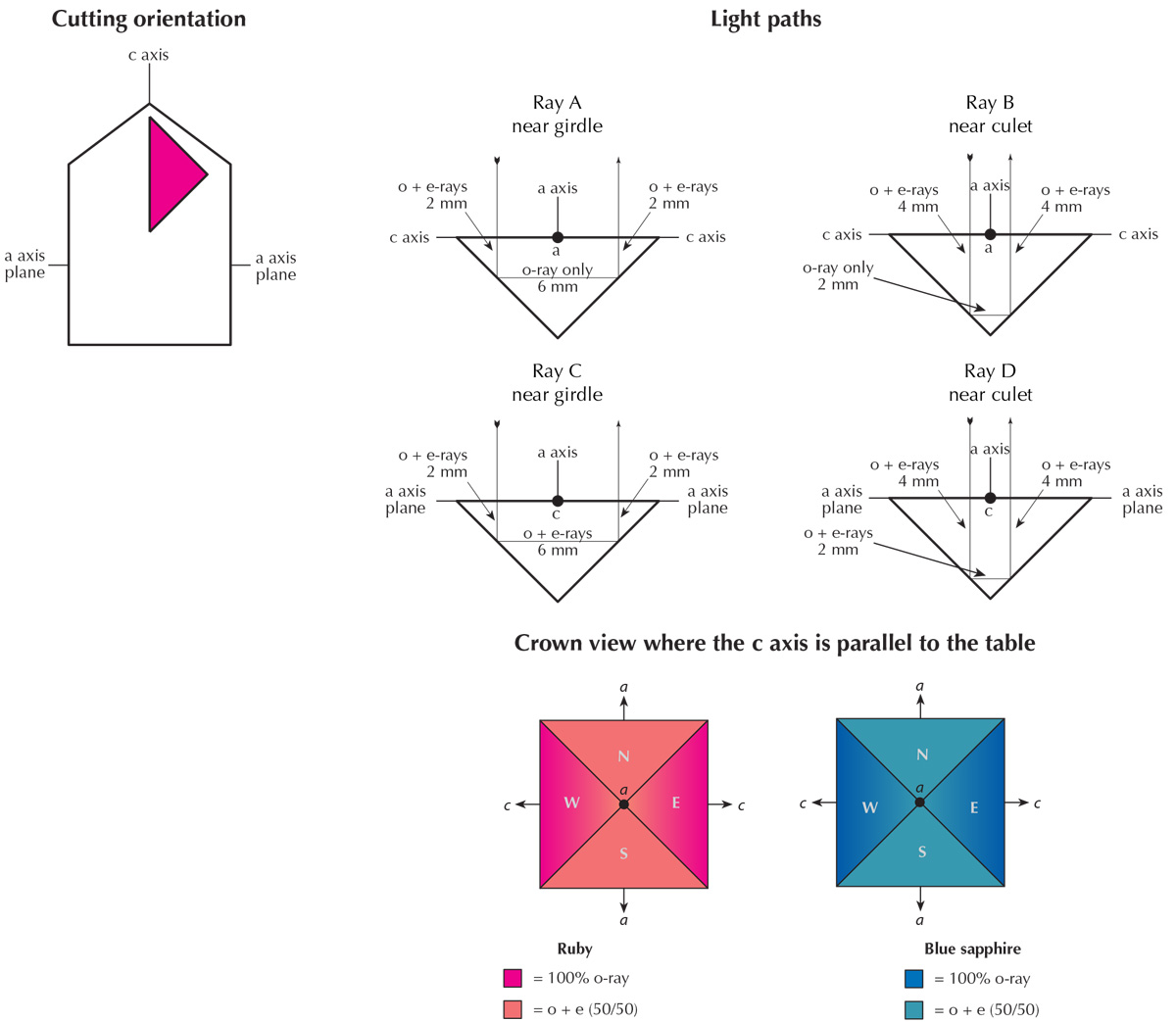 Figure 6. Pleochroism as seen through the crown in ruby and sapphire cut with the table parallel to the c axis. Illustration: Richard W. Hughes
Uniaxial with C-Axis Parallel to Table
Now let us consider a uniaxial gem cut with the c axis parallel to the table, rather than perpendicular. This is illustrated in Figure 6.
- Ray A has 6 mm of the o-ray only, and 4 mm of equally mixed o- and e-rays, giving a total of 80% o-ray and 20% e-ray.
- Ray B consists of 2 mm of o-ray only and 8 mm of equally mixed o- and e-rays, giving a total of 40% o-ray and 60% e-ray. Thus the color on these facets will show more of the o-ray near the girdle and less at the culet.
- Rays C and D are equal mixtures of o- and e-rays, regardless of light path. Thus those facets will display a uniform 50%–50% split.
In summary, when pleochroic gems are cut with the c axis parallel to the table facet, a pleochroic bow tie can be seen, as shown at the bottom of Figure 6 and in Figure 7.
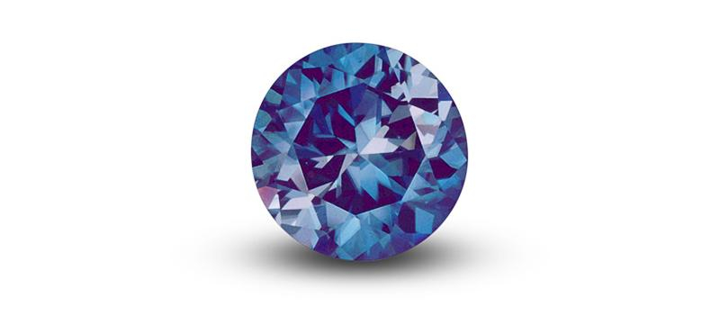 Figure 7. 15.6 ct blue zircon from Joel Arem's Color Encyclopedia of Gemstones (1977), showing an obvious pleochroic "bow tie" effect. Photo © Joel Arem
A striking example of these differences in color due to pleochroism is found in the first edition of Joel Arem 's Color Encyclopedia of Gemstones (1977). Color plate 64 shows a round 15.6 ct Cambodian blue zircon (uniaxial) that has been cut with the c axis parallel to the table facet. In blue zircon, the o-ray is blue while the e-ray is colorless. This orientation has resulted in a large white "bow tie" that is obvious in the photo. Closer examination of this photo reveals that, in the blue portion of the stone, the color is lightest at the culet and gradually deepens towards the girdle. This is because the percentage of o-ray in the color gradually increases from 50% at the culet to 100% at the girdle.
Pleochroism in Tourmaline
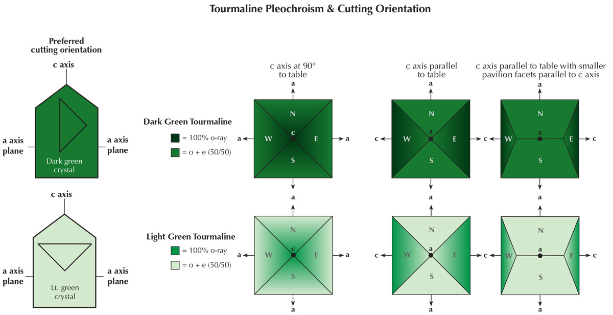 Figure 8. Pleochroism in tourmaline. Because of its strong pleochroism, dark green tourmaline (top) must be oriented to minimize the o-ray’s effect on face-up appearance. Light green tourmaline (bottom) is oriented to maximize color by increasing the o-ray's effect. Illustration: Richard W. Hughes
Tourmaline is one of the most strongly pleochroic gems. Indeed the pleochroism is so strong that it has previously been made to manufacture crude polariscopes ('tourmaline tongs'). So strong is tourmaline's pleochroism that orientation of cut stones is extremely important.
In tourmaline, the o-ray is always the darker of the two colors. Thus light-colored tourmalines will be oriented with the table perpendicular to the c axis, to get the richest color. However in dark tourmalines, the opposite orientation will be used, with the c axis parallel to the table facet. In these stones, so dark is the o-ray that the pavilion facets lying along the c axis will typically be cut very steep, to minimize the effect of the o-ray on the face-up appearance. This is shown in Figure 8.
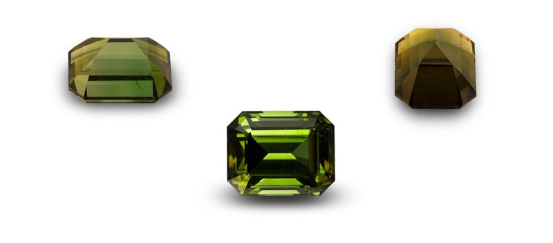 Figure 9. Pleochroism in a tourmaline cut with the c axis parallel to the table, as seen with the naked eye through the crown, side and end. Gem: Palagems.com; Photos: Wimon Manorotkul & Mia Dixon.
Pleochroic Differences In Natural & Verneuil Synthetic Corundum
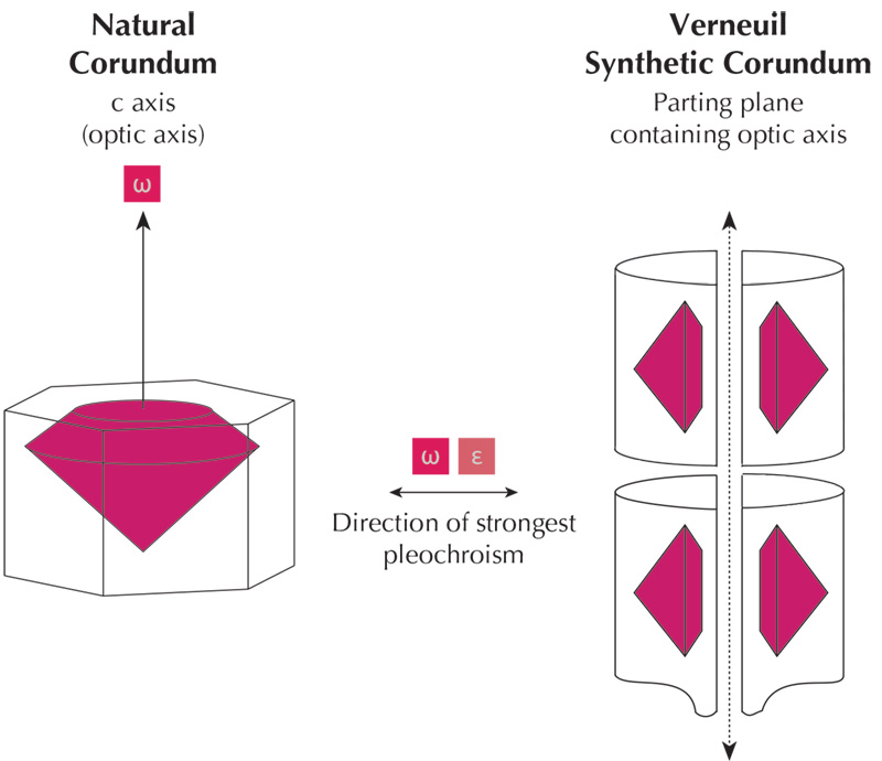 Figure 10. Orientation differences between natural and Verneuil synthetic corundum result in differences in pleochroism. Natural ruby and sapphire tends to be cut with the table facet perpendicular to the c axis. As a result, no pleochroism is visible through the table with the dichroscope. The opposite holds true for Verneuil synthetic corundum.
Differences in the pleochroism of natural and Verneuil synthetic corundum often occur because of differences in their orientation. Natural corundum is generally oriented with the c axis perpendicular to the table, which tends to provide the best color and weight retention. In the Verneuil product, the c axis always lies within the plane along which the boule is split. Though not necessarily parallel to the boule’s length, it is always parallel to the plane of the split. As most cutters of this synthetic are only concerned with weight retention—the price is the same regardless of orientation—they will normally place the table facet parallel to the split, so that the c axis lies parallel to the table. The result is that, when viewed with a dichroscope, Verneuil synthetics show dichroism through the table, whereas 80% or more of natural corundum shows little or none.
These differences are most readily apparent in blue sapphires and are least conspicuous in yellow to colorless sapphires. No pleochroism, of course, will be detected in completely colorless stones. The differences in the typical orientation of natural and Verneuil synthetic corundum are shown in Figure 10. An example of pleochroism in a Verneuil synthetic sapphire boule is shown in Figure 11.
It is important to note that in all strongly pleochroic gems cut with the optic axis or axes at oblique angles to the table, the pleochroism, if visible, will have a symmetrical pattern. The different pleochroic colors will be found 90° apart from one another, while identical pleochroic colors will lie exactly opposite each other. This is true for both uniaxial and biaxial stones, even if they are cut in a less-symmetrical (i.e. pear or heart) shape. The pleochroism will always be displayed in a symmetrical way because the important optical directions are always arranged in a symmetrical way within the crystal.
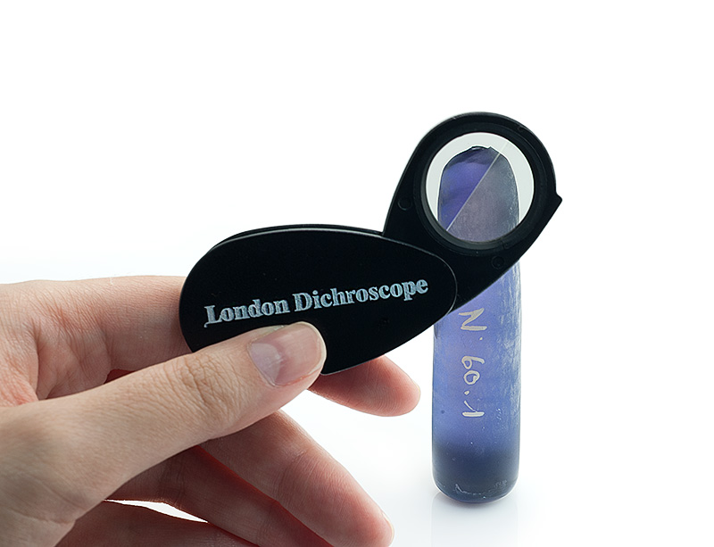 Figure 11. Pleochroism in a Verneuil-grown half-boule of synthetic sapphire, as viewed with a London (polaroid-plate) dichroscope. In Verneuil-grown synthetic corundum, the optic axis always lies in the plane of the split. Photo: Wimon Manorotkul
Pleochroism in Biaxial Gems
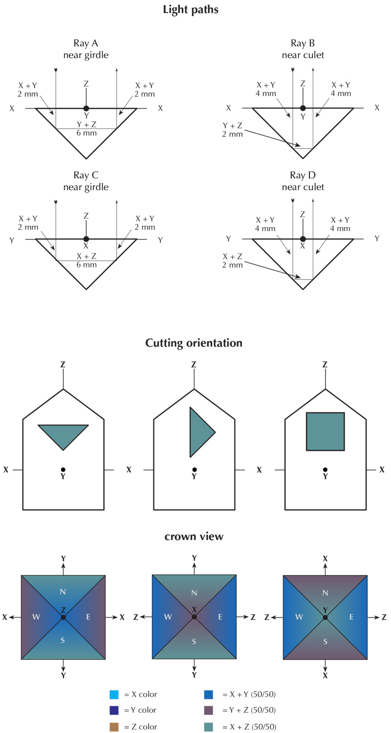 Figure 12. Pleochroism in biaxial crystals. Illustration: Richard W. Hughes
Within biaxial gems, pleochroism is displayed in a similar manner to that of uniaxial gems, except that there are three different pleochroic directions (X, Y, and Z) instead of just two.
Figure 12 shows how the pleochroism of a faceted biaxial gem might vary. Let us say that the color of the X (alpha) vibration (not travel) direction is cyan, Y (beta) is blue, and Z (gamma) is brown. The gem has been cut with Z perpendicular to the table, X running east-west, and Y running north-south.
- Ray A travels 4 mm parallel to Z, where it vibrates along X and Y, giving 20% X and 20% Y colors. It travels 6 mm parallel to X (vibrating along Y and Z), giving 30% Y and 30% Z. The total of Ray A will consist of 20% X (cyan), 50% Y (blue) and 30% Z (brown).
- Ray B travels 8 mm parallel to Z, where it vibrates along X and Y, giving 40% X and 40% Y colors. It travels 2 mm parallel to X (vibrating along Y and Z), giving 10% Y and 10% Z. The total of Ray B will consist of 40% X (cyan), 50% Y (blue) and 10% Z (brown).
- Ray C travels 4 mm parallel to Z, where it vibrates along X and Y, giving 20% X and 20% Y colors. It travels 6 mm parallel to Y (vibrating along X and Z), giving 30% X and 30% Z. The total of Ray C will consist of 50% X (cyan), 20% Y (blue) and 30% Z (brown).
- Ray D travels 8 mm parallel to Z, where it vibrates along X and Y, giving 40% X and 40% Y colors. It travels 2 mm parallel to Y (vibrating along X and Z), giving 10% X and 10% Z. The total of Ray B will consist of 50% X (cyan), 40% Y (blue) and 10% Z (brown).
In summary, we can see that biaxial stones display more complex pleochroism. Facets N and S will show different amounts of the three pleochroic colors compared with facets E and W. As with uniaxial gems, differences in color will be observed not only when comparing facets N and S to facets E and W, but also differences will be seen when comparing areas closer to or farther from the culet on the same facet. These differences are far more complex in biaxial stones, which have three unique vibration directions (as compared with only two in uniaxial stones). Keep in mind, however, that differences in color due to pleochroism will occur in a symmetrical pattern, with different pleochroic colors being 90° apart from one another, just as in uniaxial gems.
The above descriptions of pleochroism in both uniaxial and biaxial gems demonstrate the variation of color on facets of different orientations. But remember that these individual components will mix together to produce the single color seen on any individual point on each facet. A dichroscope can be used to separate that single color into its individual components.
Pleochroism, or something else?
Pleochroism is not the only factor that may cause color variations in faceted gems. Color zoning can result in significant color variations, as can different path lengths of light. Both of these attributes may be present in either singly or doubly refractive gems. Although these should not be confused with pleochroism, they sometimes are.
Consider, for example, an elongated cushion-shaped tsavorite garnet that possesses no visible color zoning as an example (Figure 13). With such a stone, the ends of the cushion will appear darker than the sides. This effect could easily be confused with pleochroism, despite the fact that pleochroism cannot occur in singly refractive stones. The actual cause of the darker end facets is the longer light paths.
The Beer-Lambert law states that absorption is directly related to the length of the light path and concentration of absorption agent in the sample. If the path length is doubled (while the amount of colorant remains uniform), then the amount of absorption is also doubled. With a 12 mm by 6 mm cushion-ct tsavorite, light from above internally reflected off the end facets travels down the entire length of the stone, nearly double the path length of light internally reflected across the side facets. As a result, the color of the end facets is nearly twice as dark as those on the side. If the same piece of rough were cut as a round, however, no variation in color would be seen. Thus the color variations sometimes seen in SR gems result from variations in light path lengths or color zoning, rather than pleochroism.
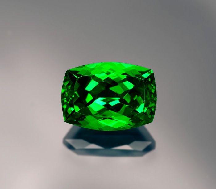 Figure 13. The color of this 7-ct tsavorite garnet is darker on the ends than in the center. This is not due to pleochroism—garnet is singly refractive—but an illustration of the Beer-Lambert law, where longer light paths result in greater absorption. Gem: Palagems.com; photo: Wimon Manorotkul
The Dichroscope
Not all doubly refractive gems show distinct pleochroism. To determine if color variations across the face of a DR gem are due to pleochroism, or simply variations in path length or color zoning (or even colored inclusions), a dichroscope should be used. Generally speaking, calcite-type dichroscopes are preferable to the polaroid types, because calcite is colorless. The polaroid filters used in polaroid dichroscopes generally have a greenish color of their own, and so may slightly modify the true pleochroic colors seen or a grayish color that diminishes light return.
In using the dichroscope, one must check uniaxial gems at right angles to the c axis (optic axis) to observe the strongest pleochroism, while biaxial gems should be examined along each of the mutually perpendicular X (alpha), Y (beta) and Z (gamma) directions. But remember that the dichroscope examines transmitted light only. Even if no pleochroism is seen while looking through the table facet with the dichroscope, pleochroic color variations may still be visible with the naked eye across the face of a faceted gem (closer to or farther from the culet). This is because the facet orientation causes light to be reflected in more than one direction as it traverses the stone, as previously described.
 Figure 14. Three tools for observing pleochroism: the calcite dichroscope, the polaroid-plate dichroscope and polaroid sunglasses. Polaroid sunglasses make an effective field tool, as simply rotating a pleochroic gem in front of them will allow one to observe the different colors (albeit one color at a time). A polariscope with the polarizer and analyzer in the parallel position can also perform the same function. Photo: Wimon Manorotkul
In DR stones, color zoning and path length variations (in non-round or non-square cut shapes) must also be taken into account in determining the existence of pleochroism.
When a faceted colored gem is being examined and described, gemologists need to recognize that the overall appearance perceived as "real" is a synthesis of many factors, some related and some not. The final determination of pleochroism (and all other color attributes) for purposes of color grading should be made with the gem in the face-up position, as that is how the stone was designed to be viewed. Yet this examination should not be restricted to a direction perpendicular to the table facet. Gems are often viewed from oblique angles, and so it seems only logical that the grading should be made in an arc ranging anywhere from parallel to the table to directions perpendicular to the table.
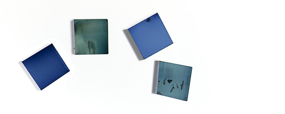
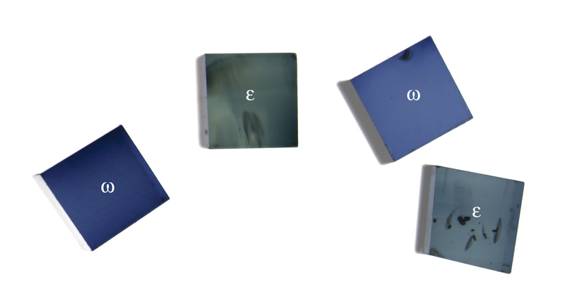 Figure 15. Small cubes of blue sapphire while viewed perpendicular to the optic axis through a polarizing filter. Rotating either the gems or polarizer will switch the two pleochroic colors of each piece. Photo: Wimon Manorotkul
Conclusion
While gemologists typically observe pleochroism with a dichroscope, or possibly a polarizing filter, this attribute's effect on appearance is more complex and generally overlooked. A basic understanding of optical crystallography unlocks the secret of faceted pleochroic gems, revealing that, when strongly pleochroic gems are examined with the naked eye, pleochroism is always present, even when viewing along an optic axis.
For some gems, pleochroism plays an important role in overall color appearance. It is the wise gemologist (and lapidary) who understands that pleochroism is far more than just the twin colors seen in a dichroscope.
|
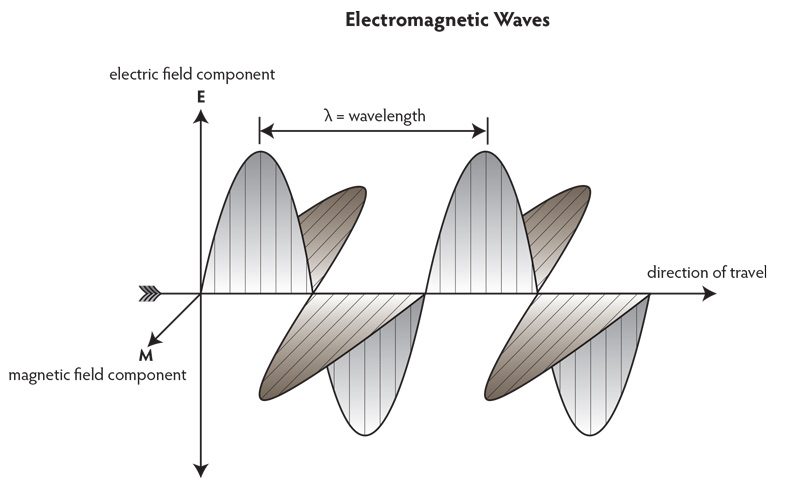
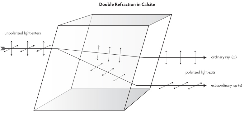
Appendix – Electromagnetic Radiation and Crystals Visible light is one form of electromagnetic radiation. Light is sometimes described as behaving like a particle, but when dealing with transmission and reflection, it is more convenient to describe light as a wave (Frye, 1974). This is often referred to as the “dual nature of light.” For the purposes of pleochroism, the wave theory is more appropriate. Electromagnetic waves consist of electric (E) and magnetic (M) portions, each of which is mutually perpendicular. The electric portion of the wave interacts with, and is altered by electrons, and so it is only the E portion of the wave that will be considered when referring to refraction and polarization (Figure A). The electric vector (vibration) of ordinary light, such as sunlight, may extend in any direction perpendicular to the direction of travel (Figure B). Such light is said to be unpolarized. Should the light wave be forced to vibrate in a single plane, the ray is polarized. When light enters a substance of greater optical density, it is slowed because the electron density is greater and thus there is greater interference with the E portion of the wave. If the electron density of that substance is random, such as in a gas or glass, the polarization is not affected. The same holds true when it enters isometric (cubic) crystals, where the lattice points are arranged identically in each of the three dimensions. In all other crystals, the lattice points (and thus the electron clouds) are unequally spaced. The spacing of lattice points in dimetric crystals varies in two dimensions, while that of trimetric crystals varies in all three dimensions. This affects the manner in which light moves through them. Non-cubic crystals are termed anisotropic. They both split and perpendicularly polarize light upon entry, except in directions where the electron clouds are symmetrical. Those directions are termed optic axes and light travels along these directions as it does in isotropic materials. When light enters an anisotropic substance in a direction not parallel to an optic axis, it splits into two segments, each of which is polarized perpendicular to the other. Polarization can also occur when light strikes a highly reflective surface, such as a lake, producing what our eyes perceive as glare. The direction of polarization is parallel to the reflective surface. If one dons “Polaroid” sunglasses, the glare is eliminated because the lenses contain tiny polarizers oriented perpendicular to the horizon.
|
![]()
About the author
Richard W. Hughes is one of the world’s foremost experts on ruby and sapphire. The author of many books and over 170 articles, his writings and photographs have appeared in a diverse range of publications, and he has received numerous industry awards. Co-winner of the 2004 Edward J. Gübelin Most Valuable Article Award from Gems & Gemology magazine, the following year he was awarded a Richard T. Liddicoat Journalism Award from the American Gem Society. In 2010, he received the Antonio C. Bonanno Award for Excellence in Gemology from the Accredited Gemologists Association. The Association Française de Gemmologie (AFG) in 2013 named Richard as one of the Fifty most important figures that have shaped the history of gems since antiquity. In 2016, Richard was awarded a visiting professorship at Shanghai's Tongji University. 2017 saw the publication of Richard and his wife and daughter's Ruby & Sapphire • A Gemologist's Guide, arguably the most complete book ever published on a single gem species and the culmination of four decades of work in gemology. In 2018, Richard was named Photographer of the Year by the Gem-A, recognizing his photo of a jade-trading market in China, while in 2020, he was elected to the board of directors of the Accredited Gemologists Association and was appointed to the editorial review board of Gems & Gemology and The Australian Gemmologist magazine. In 2022, Richard published Jade • A Gemologist's Guide, while 2024 brought Broken Bangle • The Blunder-Besmirched History of Jade Nomenclature. His jade trilogy was completed in 2025 with his translation of Heinrich Fischer's Nephrite and Jadeite.
Acknowledgments
The author would like to thank John Emmett for carefully reviewing the manuscript and Joel Arem (joelarem.com) for allowing the use of his blue zircon photo.
Notes
Based on a piece first published in Gemological Digest:
- Hughes, R.W. (1988) Pleochroism and colored stone grading. Gemological Digest, Vol. 2, No. 3, pp. 16–24.
This version is significantly edited and completely updated with new illustrations and has been published in Gems & Gemology, 2014, Vol. 50, No. 3, Fall, pp. 216–226.
For more on crystal optics, see: Hughes, R.W. (2017) Crystal optics • Gem and crystal optical properties. LotusGemology.com
References
- Arem, Joel (1977) Color Encyclopedia Of Gemstones. 1st ed. Van Nostrand Reinhold Co., New York, 147 pp.
- Frye K. (1974) Modern Mineralogy. Prentice-Hall, Englewood Cliffs, NJ, 325 pp.
- Hughes R.W. (1997) Ruby & Sapphire. RWH Publishing. Boulder, CO, pp. 80–85.
- Hurlbut, C.S. (1984) The jeweler's refractometer as a mineralogical tool. American Mineralogist, Vol. 69, pp. 391–398.