There are many benefits to using a long-wave UV torch when testing ruby and sapphire. Here are a few examples.
UV fluorescence is one of the most underrated tools used in gemological laboratories. Standard long-wave/shortwave units as well as long-wave torches are fairy affordable and accessible yet have many uses. This author has previously discussed some of these techniques. For example, the shortwave lamp can be used as an unconventional means of viewing curved striae in synthetic corundum (Hughes, E.B., 2019). The long-wave torch can be a quick and helpful way of detecting fillers in emeralds (Hughes, E.B., 2020).
Although emeralds may be the most well-known gemstone that undergoes fracture filling treatment, it is far from the only one. In our laboratory, we have come across fillers in a variety of gems, including tourmaline, tanzanite (Hughes, R.W., 2020), spinel (Hughes, R.W., 2020), and ruby and sapphire. While glass filling is a fairly well-known treatment in corundum, the presence of oils and resins as a filler is perhaps less recognized, yet also important. One technique that can help detect fillers is the use of a long-wave UV torch. Recently a few examples of its utility have come across our desks in the laboratory.
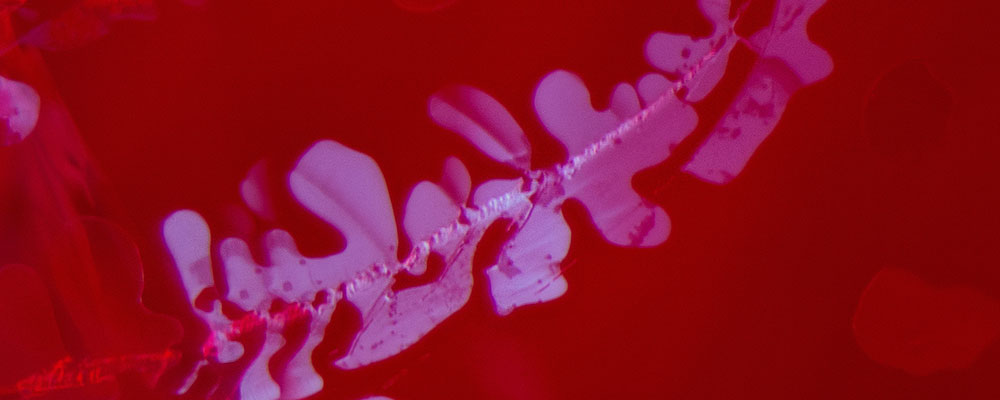
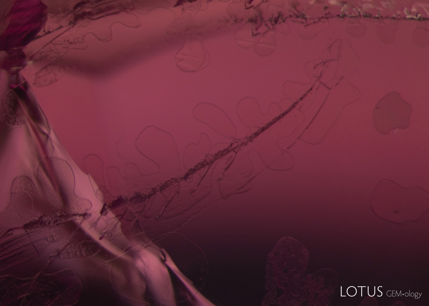
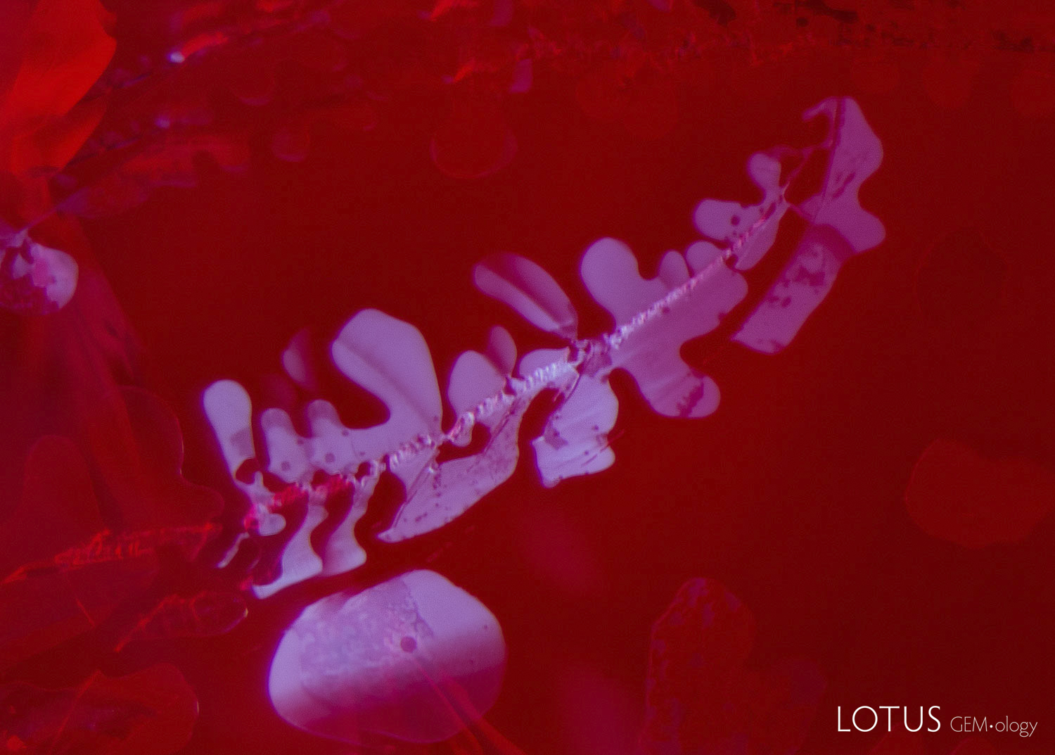
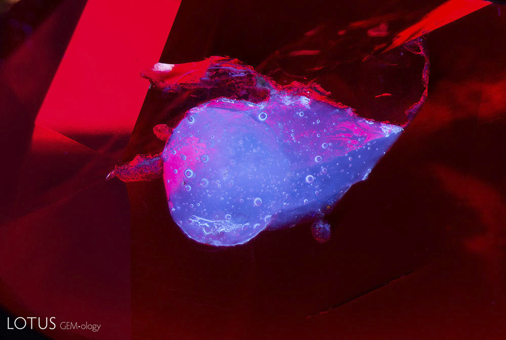
For instance, a ruby came in for testing that had several fissures. In diffuse transmitted light, a faintly dendritic pattern could be seen in the fissures. With the addition of a long-wave UV torch, the dendritic patterns lit up with a chalky fluorescent reaction that was in sharp contrast to the red fluorescence of the ruby (figure 1). Close observation confirmed that the stone was glass filled.
|
A. |
B. |
| Figure 1. A: When viewed in diffuse transmitted light, the fissure in this glass filled ruby shows a faint dendritic pattern. B: The use of the long-wave UV torch illuminates the glass filling in the fissure, so the dendritic pattern is much more visible. The chalky appearance of the filler stands out against the red fluorescence of the ruby. Photomicrographs by E. Billie Hughes/Lotus Gemology; field of view 2.5 mm. |
|
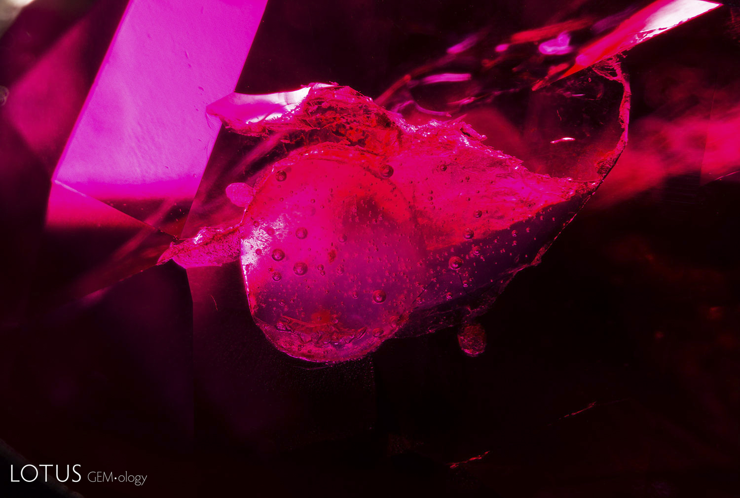
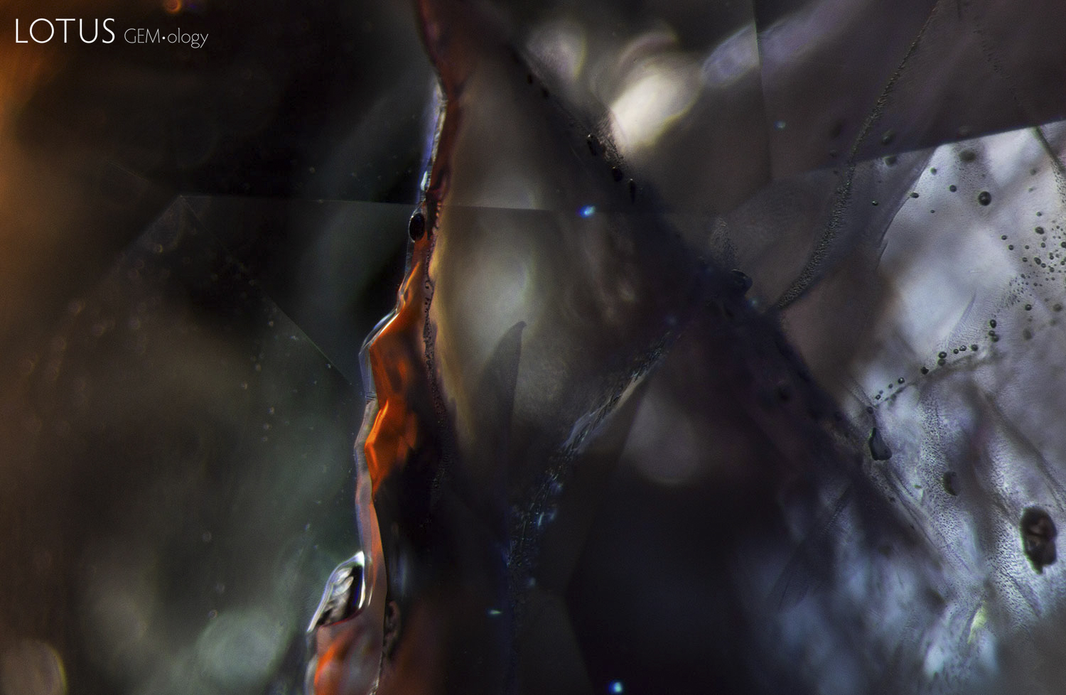
In another ruby, a surface-reaching cavity was present, which had been filled with a hardened resin, visible in dark field illumination (figure 2). However, when using a long-wave UV torch, the filler became more obvious because of its chalky appearance. The gas bubbles present were also easier to see.
|
A. |
B. |
| Figure 2. A: This ruby contains a surface reaching cavity that appears to contain hardened resin, depicted here with dark field illumination. B: By adding illumination from the long-wave UV torch, we can see the filler more clearly. Using the torch also makes the appearance of the gas bubbles more prominent. Photomicrographs by E. Billie Hughes/Lotus Gemology; field of view 7 mm. |
|
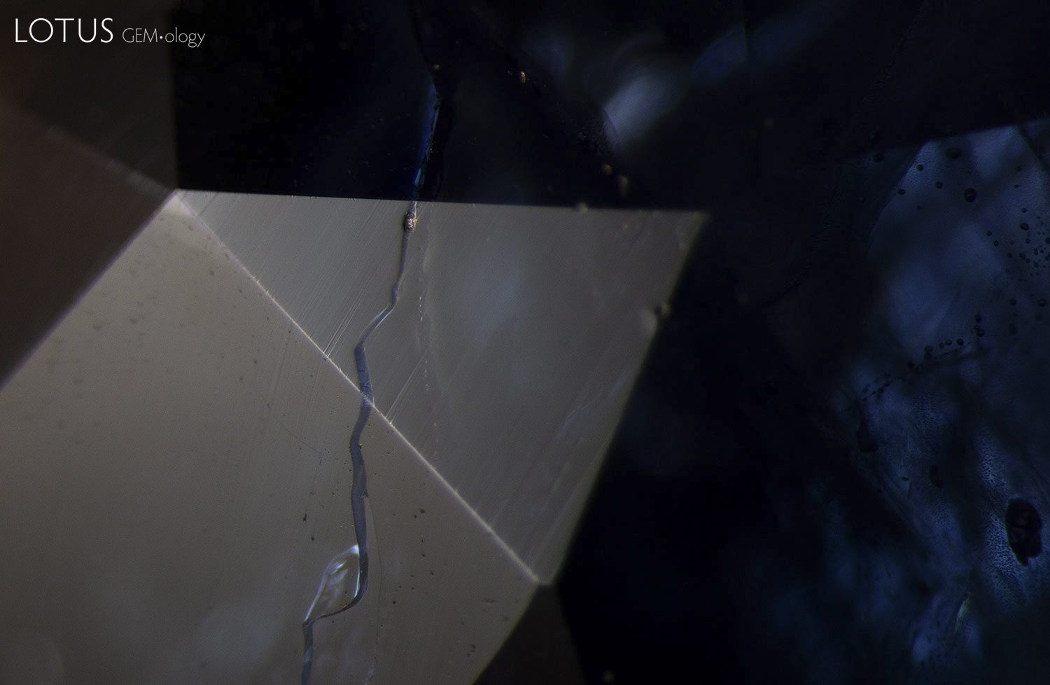
This technique is helpful not only when testing ruby, but also when testing sapphire. A blue sapphire that was submitted for testing was found to have surface reaching fissures. Observation with diffuse fiber optic light, reflected off the surface, showed a substance in the fissures with a lower luster than that of the surrounding corundum, a clear sign that the stone had been glass filled. Once long-wave UV torch illumination was added, the fissure displayed a bright orange appearance where glass was present (figure 3). In cases where glass filling is small or in very fine fissures, the bright fluorescence could be easier to see than tiny amounts of glass on the surface.
|
A. |
B. |
| Figure 3. A: Using a diffuse reflected fiber optic light, a fissure can be observed in this sapphire. Note the lower luster on the surface of the fissure, a sign of glass filling. While it is important to examine the surface with reflected light to detect glass filling, sometimes this can be hard to see if the fissure is very fine. B: The same fissure, now illuminated with the long-wave UV torch, glows a bright orange. The contrast in color makes it much easier to detect. Photomicrographs by E. Billie Hughes/Lotus Gemology; field of view 4 mm. |
|
While many of the more advanced techniques used in laboratories today can be cost prohibitive, UV torches are easily accessible. There are many affordable units available on the market. We have found the Convoy S2 6-watt model, which was used for the photomicrographs featured here, to be particularly powerful.
When testing gems, it is important to use a variety of lighting conditions, as different techniques provide different pieces of evidence. Recent experience in our laboratory demonstrates the benefits of including the long-wave UV torch in your testing procedures.
![]()
About the Authors
E. Billie Hughes is Co-Founder and Managing Director of Lotus Gemology. She oversees the company's day-to-day operations while continuing gemological research and laboratory work. After graduating from UCLA in 2011, Billie became a Fellow of the Gemmological Association of Great Britain (FGA) in 2013. Her research focuses on ruby and sapphire, including low-temperature heat treatment, and she has authored and co-authored articles in leading gemological journals. An accomplished field gemologist, she has traveled to gem deposits around the world, including nearly every major ruby and sapphire locality.
Billie is an internationally recognized educator who has lectured for trade organizations, museums, and luxury jewelry houses. She has collaborated extensively with Van Cleef & Arpels on educational programs and lectures. An award-winning photographer and photomicrographer, her images have received honors in the Nikon Small World and Gem-A competitions and have appeared in publications including National Geographic and Forbes. She is also the creator of Hyperion, Lotus Gemology's online inclusion database, reflecting her commitment to making gemological knowledge more accessible.
Billie developed an interest in gemstones from an early age, accompanying her parents on expeditions to mines and gem-producing regions around the world. That lifelong passion for fieldwork, laboratory research, education, and photography continues to shape her work at Lotus Gemology today.
Notes
This article first appeared in The Australian Gemmologist (Vol. 28, No. 3, January–June 2023, pp. 150–151).
References
- Hughes, E.B.(2019) G&G Micro-World: Curved banding in flame-fusion synthetic sapphires. Gems & Gemology, Vol.55, No. 2, Summer, pp. 264–265.
- Hughes, E.B.(2020) Long-wave ultraviolet torches • A gemmologist's new best friend. Journal of the Gemmological Association of Hong Kong, Vol. 41, pp. 57–59.
- Hughes, R.W. (2020) Gem News International: Oiled spinel. Gems & Gemology, Vol. 56, No. 3, Fall, pp. 443–444.
- Hughes, R.W. (2020) Gem News International: Oiled tanzanite. Gems & Gemology, Vol. 56, No. 4, Fall, pp. 552–553.
![]()