In our social media polls, 87% of Instagram respondents and 96% of Facebook respondents thought this ruby was unheated.
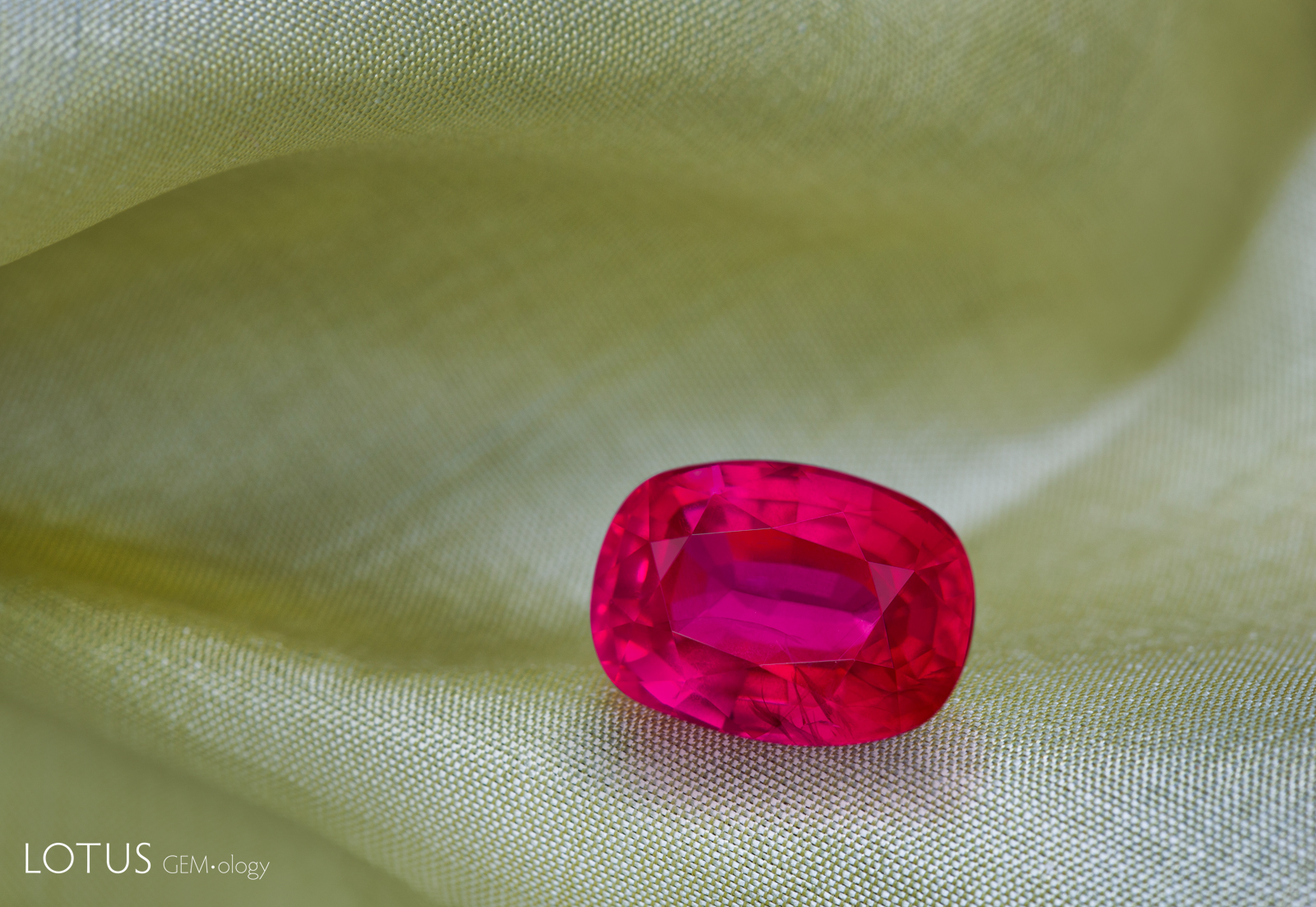 Figure 1. A 5.47-ct ruby submitted by a client for testing. Photo by Ronnakorn Manorotkul/Lotus Gemology.
It is a capital mistake to theorize before one has data. Insensibly one begins to twist facts to suit theories, instead of theories to suit facts. Arthur Conan Doyle, “A Scandal in Bohemia”
Heat Treated Burmese Ruby in Hiding
When we posted an inclusion photograph of a 5.47-ct ruby on social media and invited followers to guess whether it had been treated, the stone was widely judged to be untreated. However, laboratory testing revealed features consistent with low-temperature heat treatment, including partially dissolved rutile silk, a melted fingerprint inclusion, a 3232 cm–1;infrared absorption peak, and zoned chalky fluorescence under shortwave UV. While the appearance of silk needles alone may seem subtle and inconclusive, the combined evidence provides strong support for heating. This case highlights the limitations of image-based judgments and underscores the importance of comprehensive testing and expert interpretation in reaching an accurate conclusion.
Introduction
As gemologists, we are trained to draw conclusions based on the cumulative weight of multiple, carefully considered pieces of evidence gathered through testing. This approach stands in contrast to some contemporary discourse, particularly on social media, where definitive conclusions are often drawn from a single image. It is now commonplace to see a photograph shared online accompanied by requests such as, “Can anyone help identify this stone? Is it heated?” The ensuing responses frequently range from confident declarations of synthetic origin to equally assured claims of natural, untreated status.
Our laboratory regularly receives similar inquiries. Requests to determine treatment or origin based solely on emailed photographs, often citing the inconvenience of submitting the stone for examination, are unfortunately ones we must decline. While such questions are understandable, they underscore a fundamental principle of our work: reliable conclusions cannot be reached without appropriate testing.
A 5.47-ct ruby recently submitted to our laboratory provided a particularly clear illustration of the risks inherent in issuing conclusions without in-person analysis.
Gemological testing revealed that it was a heated ruby with characteristics that were a good match for Myanmar (Burma). While evidence clearly demonstrated that the stone was heated, it was not likely at high temperature. Clouds of rutile silk needles were present, and while in areas the needles had started to dissolve and develop small gaps, much of the silk was still visible and intact (Figure 2).
We occasionally post gemological trivia questions on social media to provide fun educational content. When we posted an inclusion photograph of this ruby (Figure 2) to invite our followers to guess whether it was treated, the response was striking. Based solely on this image, the overwhelming majority of respondents thought the ruby was untreated. A total of 87% of respondents on Instagram and 96% on Facebook guessed that it was unheated. The reveal that the stone was heated surprised most participants. Several followers who had guessed the stone was unheated requested further explanation of how this conclusion was reached. This highlights a critical point. Judgments based on a single image can be misleading when they are not supported by additional gemological evidence. In the following sections, we present the evidence that led to the conclusion that this stone was heated.
Microscopy
The first line of evidence comes from microscopic examination. In the first inclusion photograph (Figure 2), clouds of rutile silk needles can be observed.
Each inclusion can respond differently to heat treatment. Even among inclusions classified as solid, their sensitivity to heat varies. For example, primary rutile is generally less sensitive to heating than mica. Rutile silk tends to start showing heat alteration only at temperatures of approximately 1200°C or higher. (Hughes et al., 2017 and Hughes & Vertriest, 2022).
The ruby examined in this study contained silk needle inclusions. At first glance, these silk needles appear largely intact. However, closer examination reveals that many needles are partially dissolved and fragmented, which is an indicator of heat treatment. The early stage of silk breakdown suggests low-temperature heating (Hughes et al., 2017).
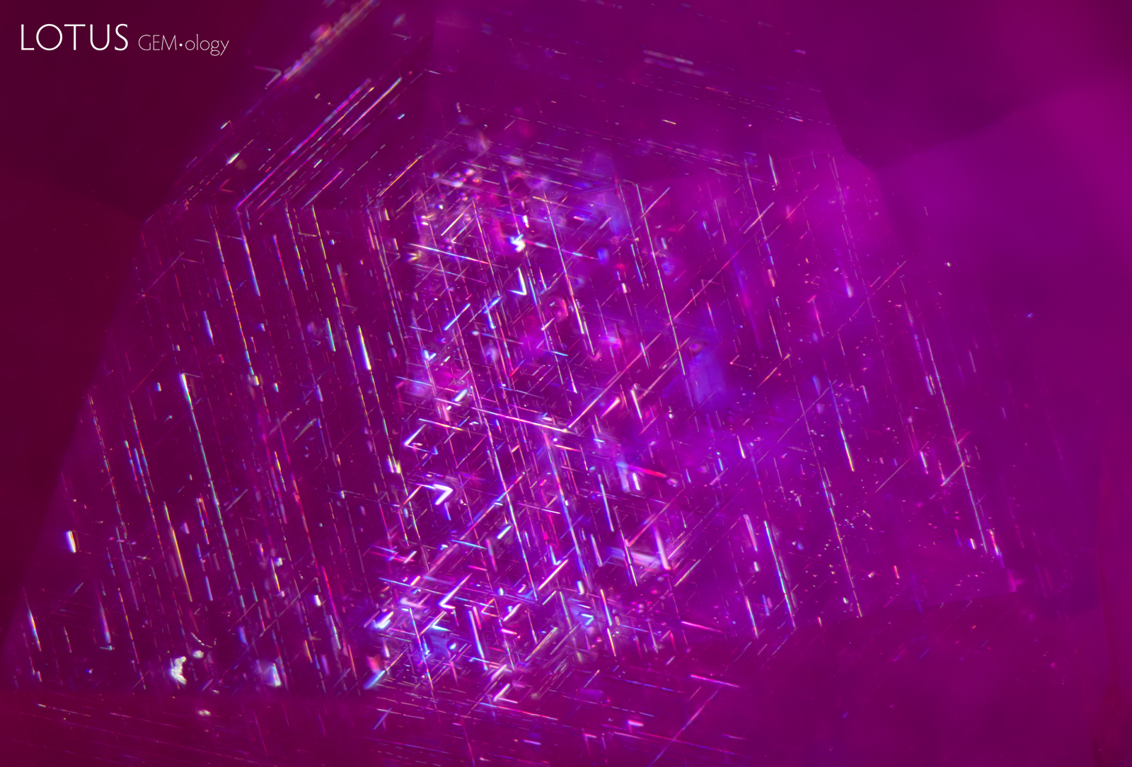 Figure 2. A cluster of rutile silk needles in a heated Burmese ruby. While superficially the rutile silk in this ruby may look intact, closer observation reveals that some of the needles have started to break down and develop gaps. Photo: E. Billie Hughes/Lotus Gemology. Field of view 3 mm.
Further evidence was revealed by changing the perspective of observation. By changing the viewing direction of the stone during microscopic examination, additional features became visible. One such feature was a melted fingerprint, which provides further support for heat treatment in this ruby (Figure 3).
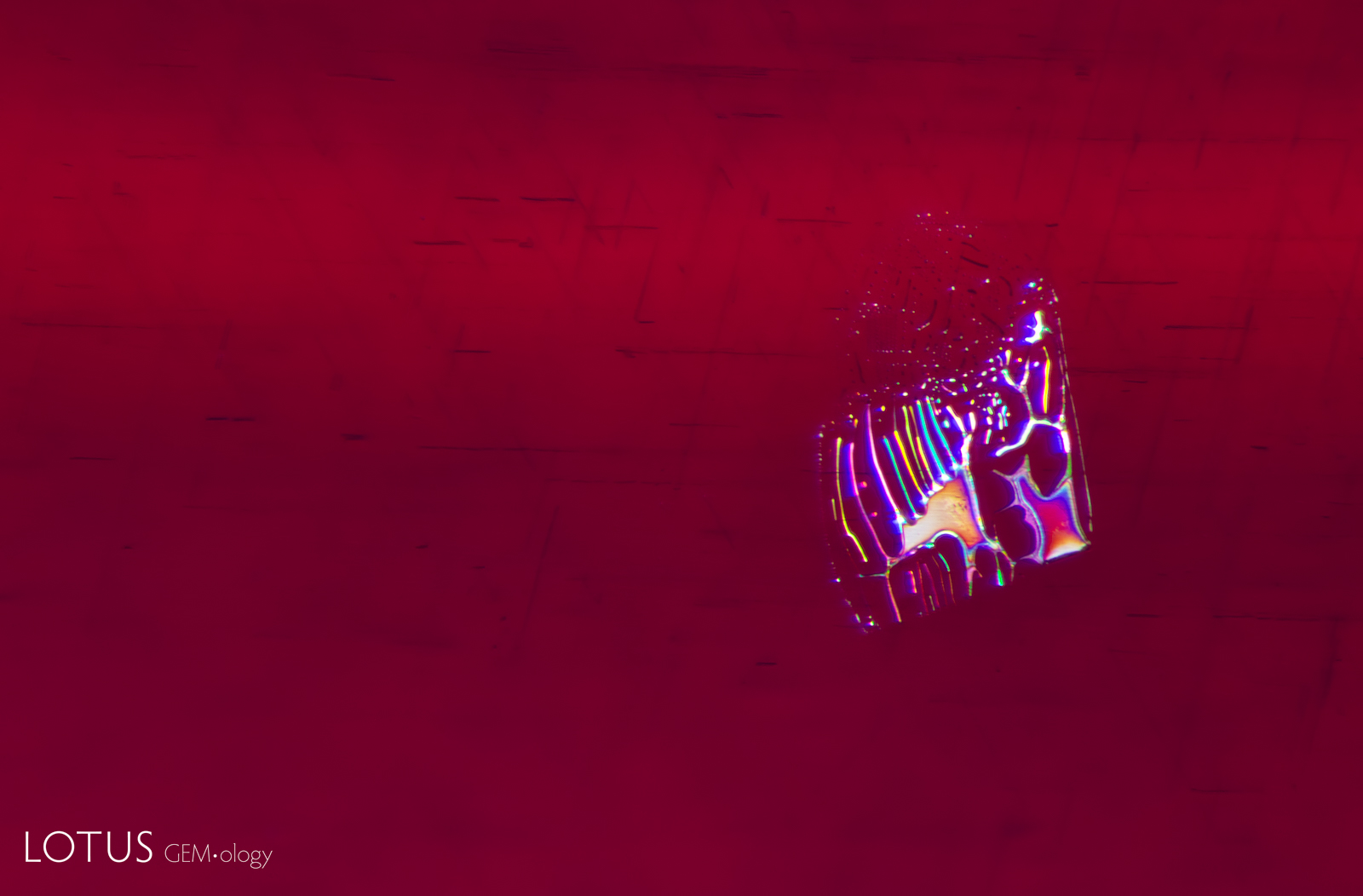 Figure 3. A melted fingerprint with drippy edges provides evidence that this ruby was heated. Photo: E. Billie Hughes/Lotus Gemology. Field of view 1.8 mm.
Infrared Spectroscopy
The next important piece of evidence comes from infrared spectroscopy. Fourier-transform infrared (FTIR) spectroscopy was conducted using a Bruker Tensor 27 spectrometer equipped with a Pike DRIFTS accessory. Spectra were collected over 256 scans at a resolution of 4 cm–1.
This ruby displays absorption peaks at 3309 and 3232 cm–1, as shown in Figure 4, which are associated with the OH stretching modes of water within the corundum structure (Breeding and Ahline, 2024). The 3232 cm–1 peak is associated with heat treatment, and is particularly useful for diagnosing low-temperature heat treatment in Mozambique rubies (Krzemnicki, 2018; Pardieu et al., 2015; Saeseaw et al., 2018) and pink sapphires from Madagascar (Saeseaw et al., 2020).
Note, however, that the absence of the 3232 cm–1 peak does not necessarily indicate that a stone is unheated (Hughes and Vertriest, 2022).
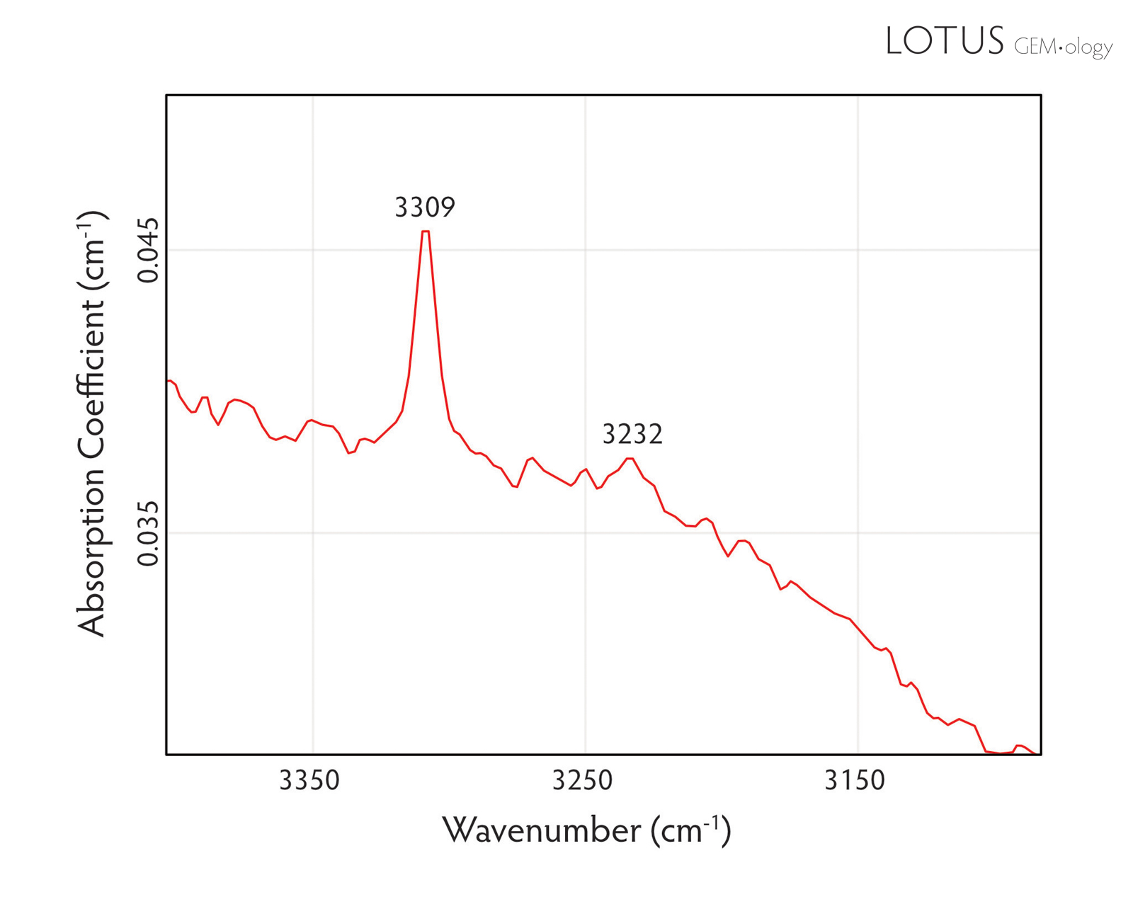 Figure 4. FTIR spectrum of a heated Myanmar ruby displaying absorption peaks at 3309 and 3232 cm–1.
Fluorescence
In addition to inclusion features and infrared spectroscopy, the final piece of evidence was obtained from observation under shortwave UV illumination. The ruby displayed red fluorescence with zoned chalky areas that correspond to the pattern of partially dissolved rutile silk (Figure 5). This is a strong indicator of heat treatment (Hughes and Vertriest, 2022).
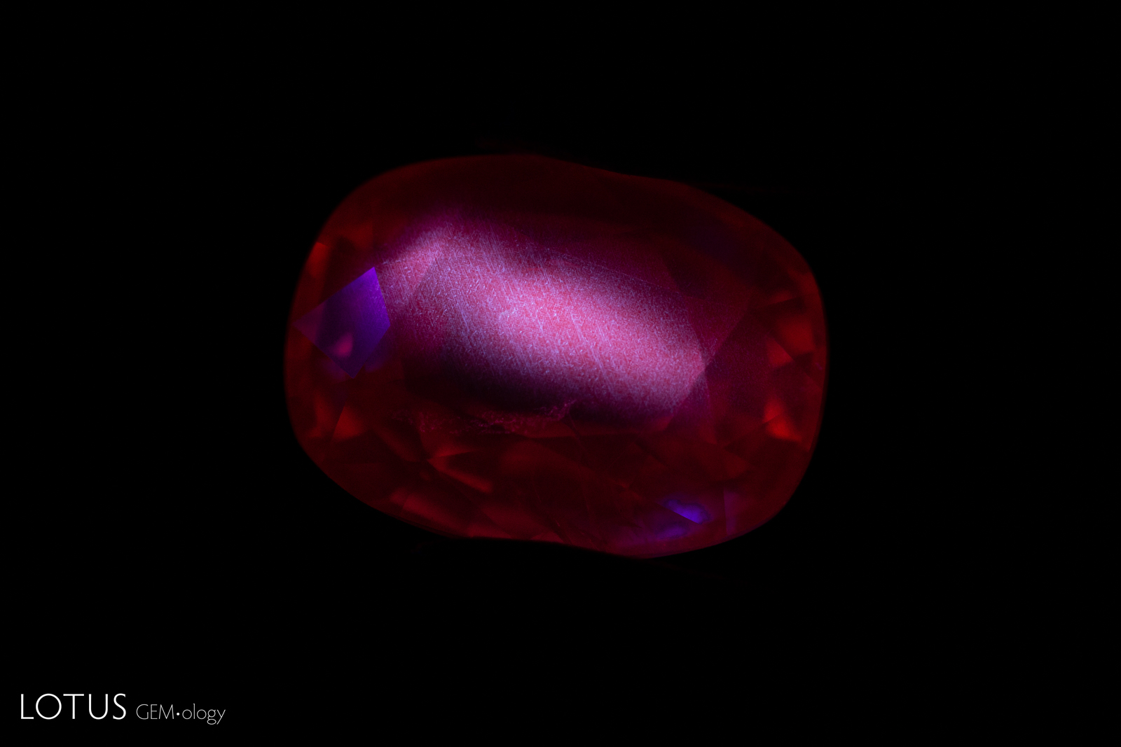 Figure 5. A heated Myanmar ruby displays red fluorescence with clear zoned chalky areas correlated with partially dissolved silk in the stone. Photo: E. Billie Hughes/Lotus Gemology.
During heat treatment, the tiny rutile silk inclusions begin to dissolve. This process releases titanium (Ti4+) ions, which then diffuse into the corundum crystal. Because titanium diffuses slowly within the crystal, it tends to accumulate in specific areas rather than being evenly distributed. At first, other ions such as iron (Fe2+) and magnesium (Mg2+) help balance the electrical charge created by the added titanium. As the titanium concentration continues to increase, aluminum vacancies form to maintain charge balance.
The interaction between titanium ions and aluminum vacancies produces the chalky fluorescence. This effect appears specifically in areas where rutile silk was originally present, because these zones become enriched in titanium after the silk dissolves, as illustrated in Figure 5. However, the absence of visible chalky fluorescence does not necessarily indicate that a ruby is unheated. Several factors can influence the appearance of this feature. In low-iron rubies, strong red fluorescence caused by chromium can mask the chalky fluorescence. In high-iron rubies, iron acts as a quencher of fluorescence. In addition, the chalky effect may be destroyed during annealing at temperatures above approximately 1500°C (D’Haenens-Johansson et al., 2025; Hughes et al., 2017).
Conclusion
In conclusion, this stone shows several features that together indicate heat treatment, including partial breakdown of rutile silk, the presence of a melted fingerprint inclusion with drippy edges, a 3232 cm–1 peak in the infrared spectrum, and zoned chalky fluorescence. Taken together, these observations illustrate the importance of evaluating multiple lines of evidence when determining treatment status, particularly in low-temperature heat-treated stones, where the effects of heating are subtle and different inclusion types respond differently to heat.
This case also highlights the limitations of judging gemstones from a single image. When a stone contains inclusions that are less sensitive to heating, heat-related changes may be difficult to recognize based on inclusion appearance alone. Without support from other gemological instruments and the expertise of a trained gemologist, such assessments can easily lead to incorrect conclusions.
Accurate conclusions therefore require consideration of all available gemological evidence and expert interpretation, rather than dependence on any single piece of evidence.

About the Authors
E. Billie Hughes is Co-Founder and Managing Director of Lotus Gemology, where she oversees the company's day-to-day operations while continuing gemological research and laboratory work. After graduating from UCLA, she became a Fellow of the Gemmological Association of Great Britain (FGA). Her research focuses on ruby and sapphire, including low-temperature heat treatment, and she has authored and co-authored articles in leading gemological journals. She has conducted fieldwork at gem deposits around the world, including nearly every major ruby and sapphire locality.
Billie is an internationally recognized educator who has lectured for trade organizations, museums, and luxury jewelry houses, including extensive educational work with Van Cleef & Arpels. She has also served as a judge on Thailand's The Secret Gems television series. An award-winning photographer and photomicrographer, her images have received honors in the Nikon Small World and Gem-A competitions and have appeared in publications including National Geographic and Forbes. She is also the creator of Hyperion, Lotus Gemology's online inclusion database.
Billie developed an interest in gemstones from an early age, accompanying her parents on expeditions to mines and gem-producing regions around the world. That lifelong passion for fieldwork, laboratory research, education, and photography continues to shape her work at Lotus Gemology today.
Natcha ('Ing') Rattana-anan has been interested in gems since childhood, which inspired her to study gemology at Chiang Mai University, where she did an award-winning thesis on heat treatment of amethyst. After graduating in 2024, she joined Lotus's gemological team.
Notes
This article first appeared in The Australian Gemmologist, 2026, Vol. 29, No. 3, pp. 221–223.
References
- Breeding, C.M. and Ahline, N.J. (2024) Infrared spectroscopy and its use in gemology. Gems & Gemology, Vol. 60, No. 4, Winter, pp. 474–492. https://doi.org/10.5741/gems.60.4.474
- D’Haenens-Johansson, U. F. S., Eaton-Magaña, S., Towbin, W. H., & Myagkaya, E. (2024). Glowing gems: Fluorescence and phosphorescence of diamonds, colored stones, and pearls. Gems & Gemology, Vol. 60, No. 4, Winter, pp. 560–580. https://doi.org/10.5741/gems.60.4.560
- Hughes, E. B., & Vertriest, W. (2022). A canary in the ruby mine: Low-temperature heat treatment experiments on Burmese ruby. Gems & Gemology, Vol. 58, No. 4, pp. 400–423. https://doi.org/10.5741/gems.58.4.400
- Hughes R.W., Manorotkul W., Hughes E.B. (2017) Ruby & Sapphire: A Gemologist’s Guide. RWH Publishing/Lotus Publishing, Bangkok, 816 pp.
- Krzemnicki, M. S. (2018). New research by SSEF studies methods for detecting low-temperature heated rubies from Mozambique. Swiss Gemmological Institute (SSEF). https://www.ssef.ch/wp-content/uploads/2018/09/SSEF-PRESS-RELEASE-New-research-by-SSEF-studies-methods-for-detecting-low-temperature-heated-rubies.pdf
- Pardieu, V., Saeseaw, S., Detroyat, S., Raynaud, V., Sangsawong, S., Bhusrisom, T., Engniwat, S., & Muyal, J. (2015). “Low-temperature” heat treatment of Mozambique ruby: Results report. GIA News from Research. https://www.gia.edu/doc/Moz_Ruby_LowHT_US.pdf
- Saeseaw, S., Kongsomart, B., Atikarnsakul, U., Khowpong, C., Vertriest, W., & Soonthorntantikul, W. (2018). Update on “low-temperature” heat treatment of Mozambican ruby: A focus on inclusions and FTIR spectroscopy. GIA News from Research. https://www.gia.edu/ongoing-research/update-low-temperature-heat-treatment-mozambican-ruby-focus-on-inclusions-and-ftir-spectroscopy
- Saeseaw, S., Khowpong, C., & Vertriest, W. (2020). Low-temperature heat treatment of pink sapphires from Ilakaka, Madagascar. Gems & Gemology, Vol. 56, No. 4, Winter, pp. 448–457. https://doi.org/10.5741/GEMS.56.4.448