Spinel Inclusions
Explore spinel inclusions in Hyperion, the Lotus Gemology inclusion database. Hyperion contains photomicrographs of inclusions and internal features in natural spinel, documented with information on geographic origin, treatment, lighting conditions, field of view, photographer and published references.
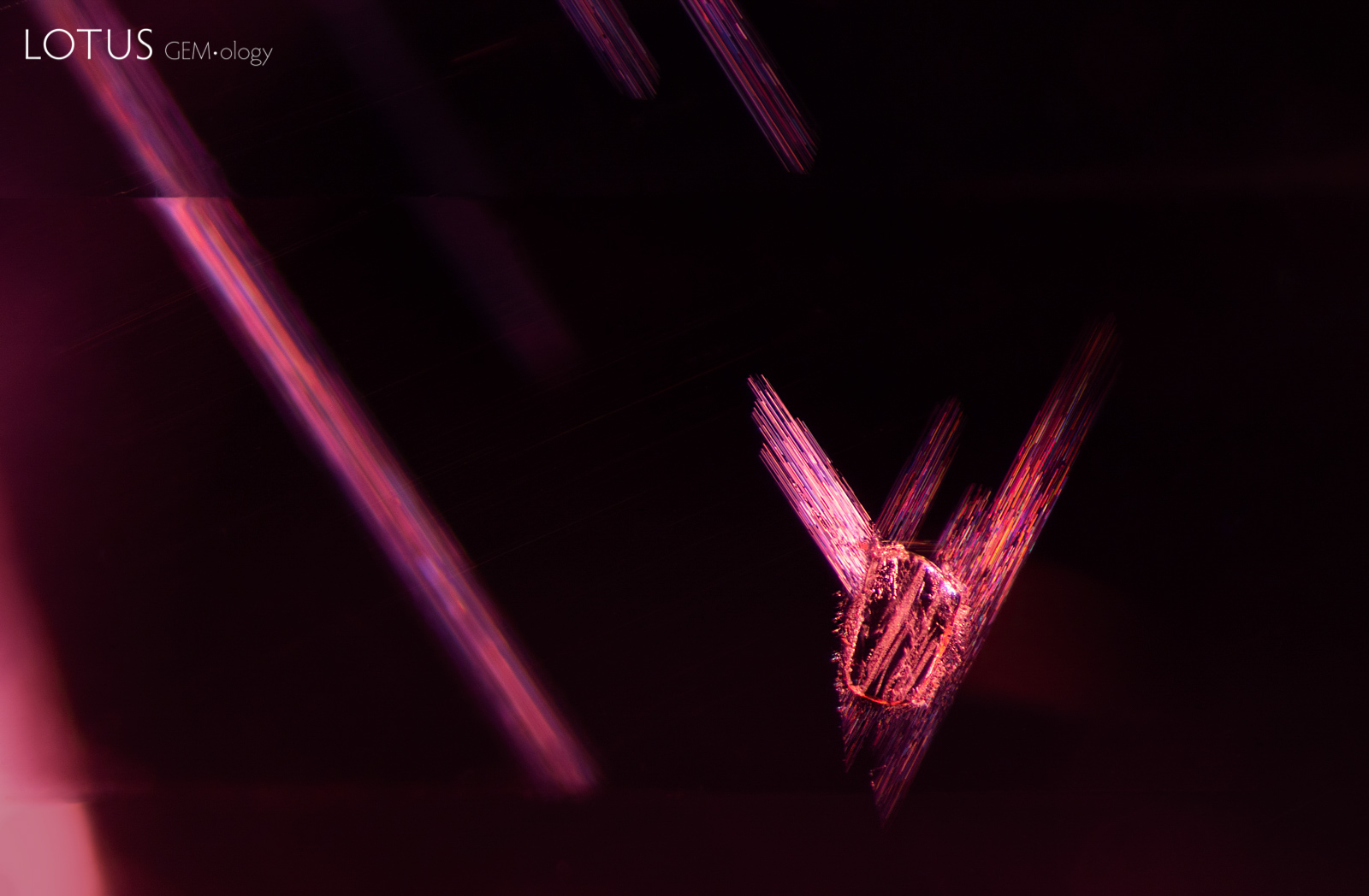
I believe I can fly. A dolomite crystal (identified with micro Raman) appears to take flight as it is flanked by dislocation needle “wings.”
Natural Spinel •
Vietnam •
Enhancements:
None Detected (None) •
Lighting Conditions:
Dark Field + Diffuse Fiber Optic
Photographer:
E. Billie Hughes •
Image Number:
A-004-1943-1
• Field of View = 4mm
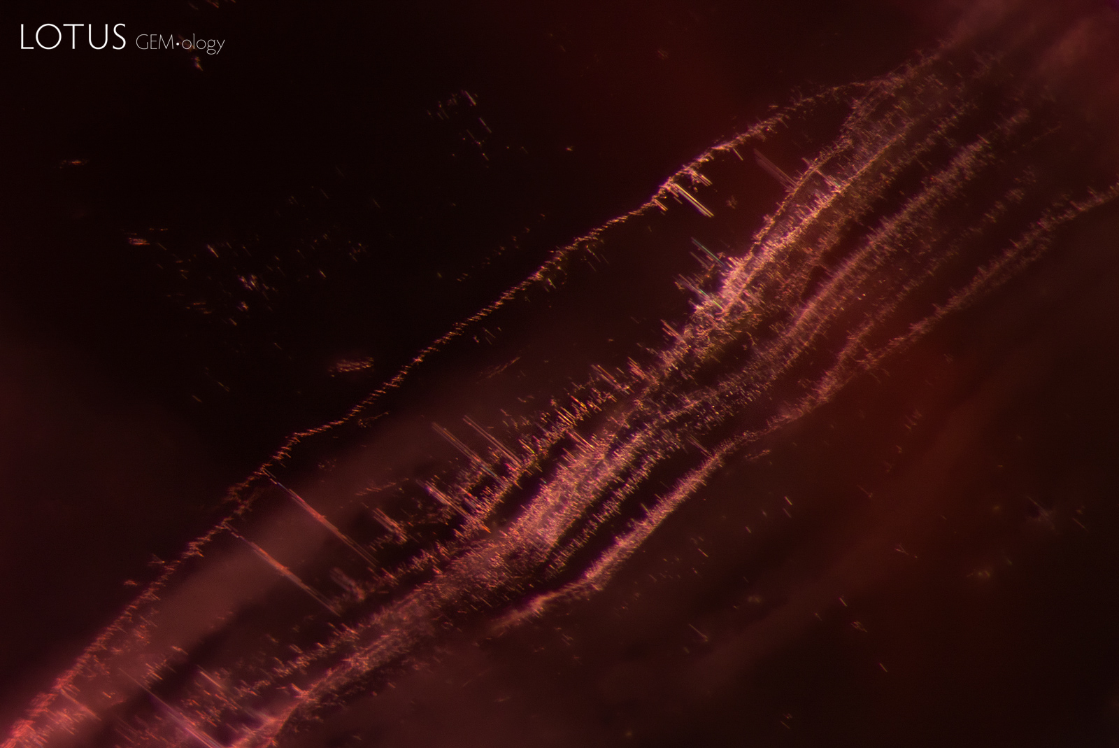
Streams of fine particles form delicate waves in this spinel. Note that the streams are made of tiny dislocation needles.
Natural Spinel •
Vietnam •
Enhancements:
None Detected (None) •
Lighting Conditions:
Dark Field
Photographer:
E. Billie Hughes •
Image Number:
A-004-1946-1
• Field of View = 3mm
A series of planes can be observed in this spinel when using fiber optic illumination. Such planes have been seen in Tanzanian spinels of various colors.
Natural Spinel •
Tanzania; Mahenge •
Enhancements:
None Detected (None) •
Lighting Conditions:
Fiber Optic: Diffuse Oblique
Photographer:
E. Billie Hughes •
Image Number:
A-004-1824-1
• Field of View = 3mm
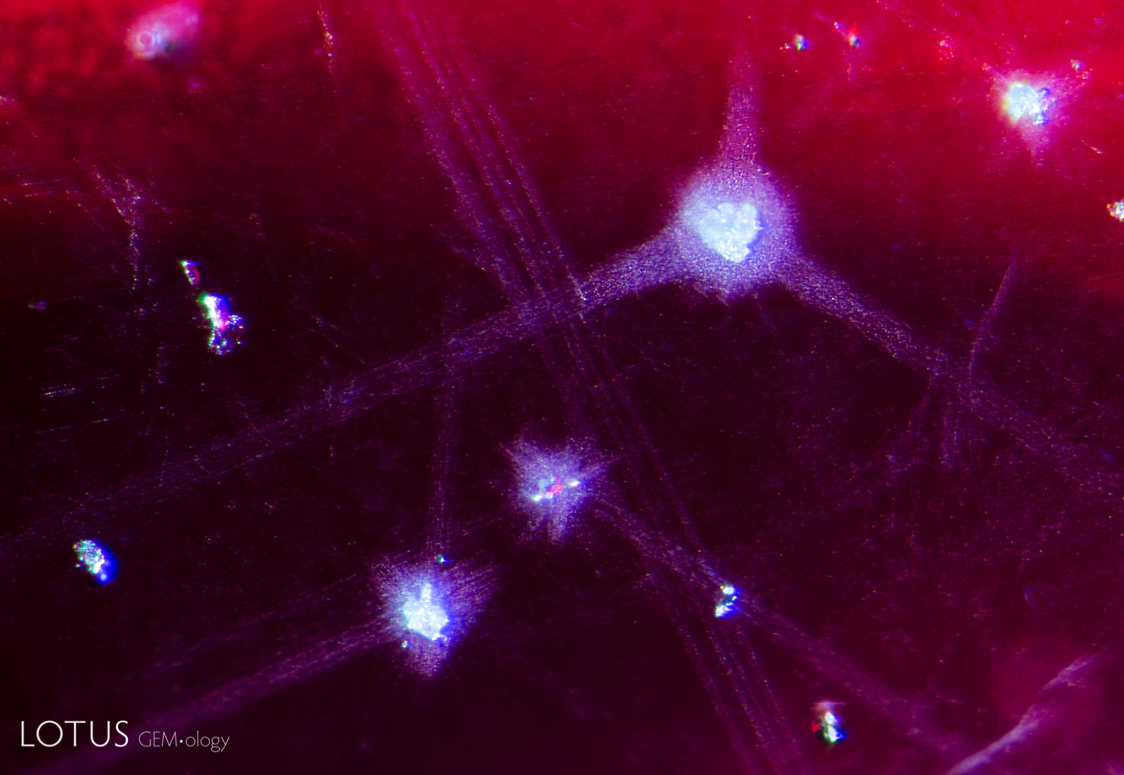
Star-like accretion halos give the interior of this spinel a galactic appearance.
Natural Spinel •
Vietnam •
Enhancements:
None Detected (None) •
Lighting Conditions:
Fiber Optic: Oblique
Photographer:
Wimon Manorotkul •
Image Number:
A-004-1769-2
• Field of View = 2.5mm
Streams of small particles create a striking pattern in this spinel from Tanzania.
Natural Spinel •
Tanzania; Mahenge •
Enhancements:
None Detected (None) •
Lighting Conditions:
Dark Field
Photographer:
E. Billie Hughes •
Image Number:
A-004-1280-2
• Field of View = 2.5mm
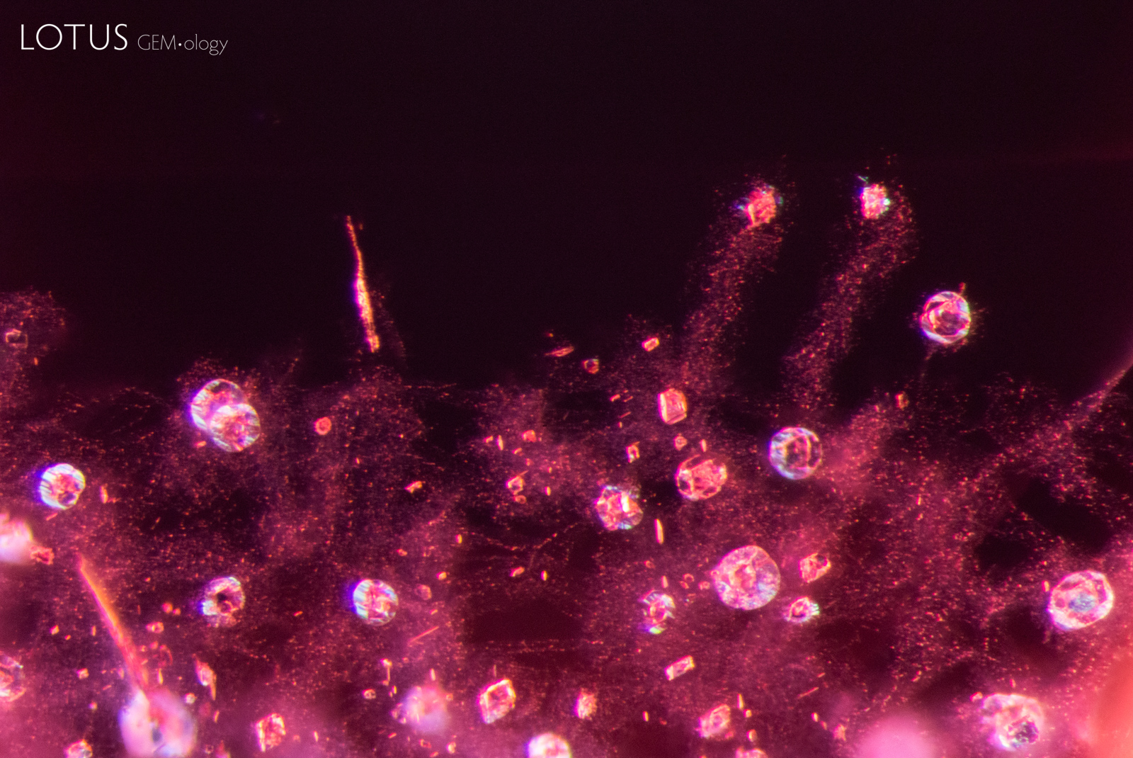
Tiny octahedral crystals, surrounded by small fissures, rest in a cloud of fine particles in this spinel from Tanzania.
Natural Spinel •
Tanzania; Mahenge •
Enhancements:
None Detected (None) •
Lighting Conditions:
Dark Field + Diffuse Fiber Optic
Photographer:
E. Billie Hughes •
Image Number:
A-004-1280-1
• Field of View = 2mm
This cluster of blocky transparent crystals was identified as apatite using Raman spectroscopy.
Natural Spinel •
Tanzania •
Enhancements:
None Detected (None) •
Lighting Conditions:
Dark Field + Diffuse Fiber Optic + Blue Filter
Photographer:
E. Billie Hughes •
Image Number:
A-004-1258-3
• Field of View = 2mm
This crystal displays multicolored iridescence on its surface when illuminated with fiber optic light. Analysis with micro Raman revealed that this crystal is phlogopite mica.
Natural Spinel •
Tanzania •
Enhancements:
None Detected (None) •
Lighting Conditions:
Dark Field + Diffuse Fiber Optic + Blue Filter
Photographer:
E. Billie Hughes •
Image Number:
A-004-1258-2
• Field of View = 3mm
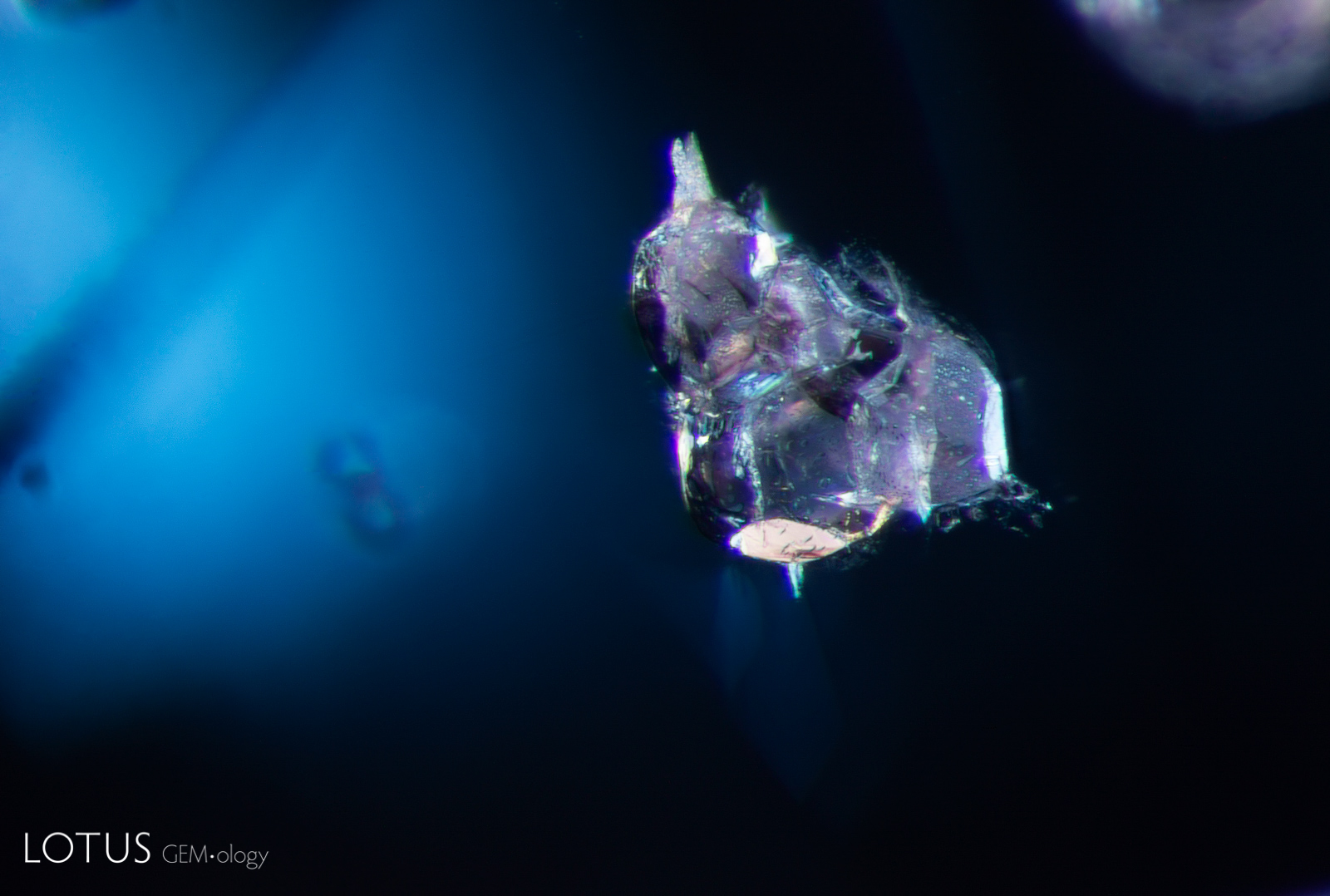
Dolomite crystals, identified with micro Raman, stand out in this blue spinel.
Natural Spinel •
Tanzania •
Enhancements:
None Detected (None) •
Lighting Conditions:
Dark Field + Diffuse Fiber Optic + Blue Filter
Photographer:
E. Billie Hughes •
Image Number:
A-004-1258-1
• Field of View = 3mm
Ghost planes light up like a multicolored rain storm when viewed between crossed polars in this cobalt blue spinel from Tanzania.
Natural Spinel •
Tanzania •
Enhancements:
None Detected (None) •
Lighting Conditions:
Light Field + Crossed Polars
Photographer:
Richard W. Hughes •
Image Number:
A-004-1078-1
• Field of View = 3mm
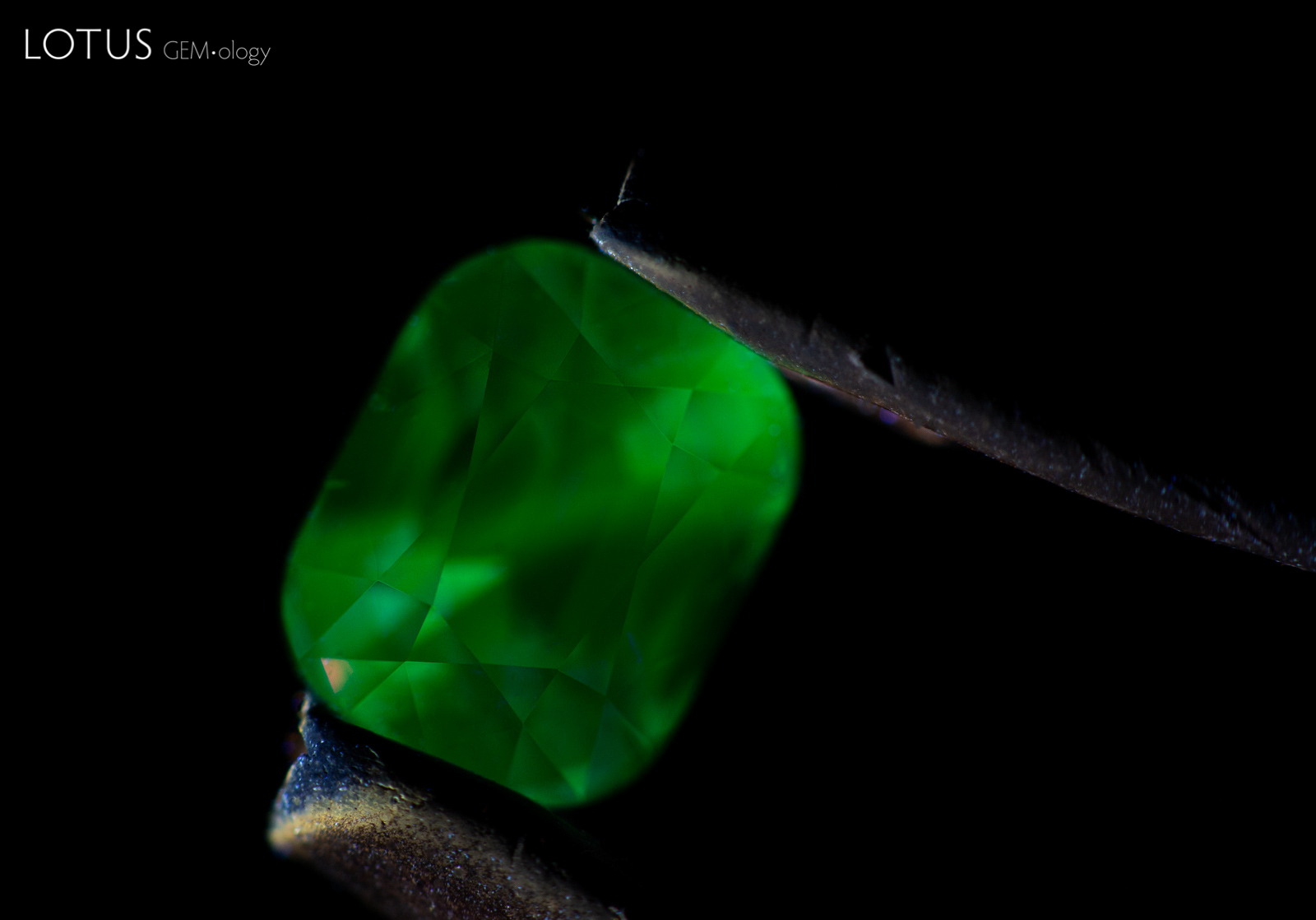
This blue spinel displayed a striking green fluorescence when illuminated with longwave ultraviolet light.
Natural Spinel •
Vietnam •
Enhancements:
None Detected (None) •
Lighting Conditions:
Ultraviolet: Longwave
Photographer:
E. Billie Hughes •
Image Number:
A-004-1033-2
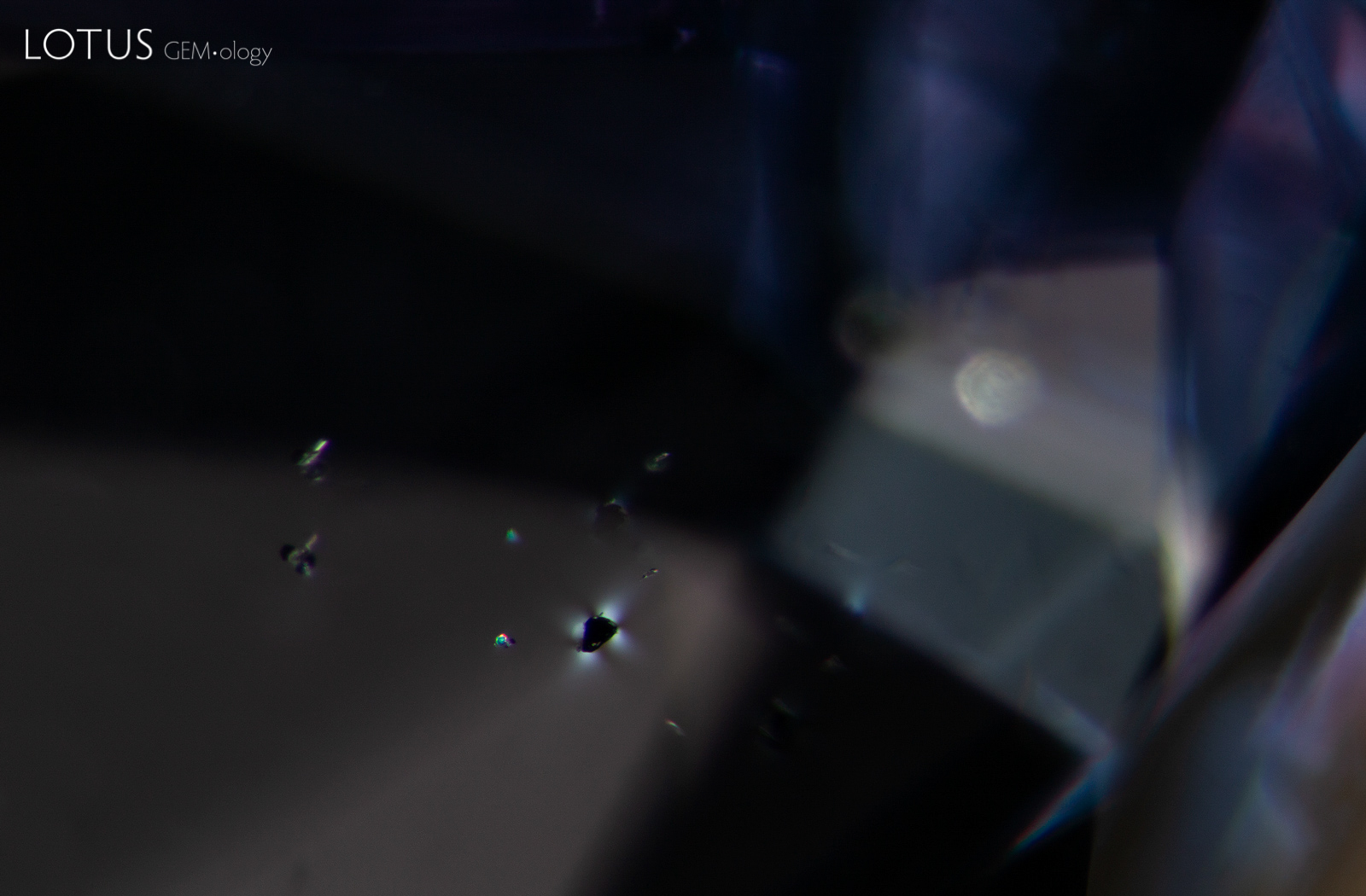
A small black crystal, likely uraninite, displays a strain pattern in crossed polars.
Natural Spinel •
Vietnam •
Enhancements:
None Detected (None) •
Lighting Conditions:
Dark Field + Crossed Polars
Photographer:
E. Billie Hughes •
Image Number:
A-004-1033-1
• Field of View = 3mm
Search the Complete Hyperion Database
Spinel inclusions may include mineral crystals, healed fissures, fluid inclusions, negative crystals, growth features and other internal features formed during or after crystal growth. Such features are important to gemologists because they can provide evidence useful in identifying spinel, understanding its geological history, recognizing treatment and, in some cases, determining geographic origin.
Spinel Inclusion Photomicrographs
The Hyperion spinel gallery includes microscopic features found in spinels from major deposits around the world. Each entry is accompanied by descriptive information and, where available, references to the gemological literature.
What Inclusions Can Reveal About Spinel
Inclusions are an important part of spinel identification. Their form, composition and alteration can help gemologists distinguish natural spinel from synthetic material, recognize evidence of treatment, and study the geological environment in which the spinel formed.
Hyperion combines inclusion photomicrography with supporting gemological information so that individual features can be compared with documented examples from known spinel deposits.
About Hyperion
Hyperion is the Lotus Gemology searchable inclusion database. It allows users to browse gemstone inclusions by gem type, geographic origin, treatment and keyword.