Gemstone Inclusions Bibliography

Gemstone Inclusions Bibliography

Explore books, articles and historical sources on inclusions in gemstones in Four Treasures, the Lotus Gemology reference database. This bibliography brings together matching records from the collection, with publication details, abstracts and cover images where available.

Gemstone Inclusion References

The list below matches the terms inclusion or inclusions in the database's searchable fields. Some references discuss inclusions as part of a broader subject.
541–560 of 1,247 matching references

Gem Notes: Blue gahnite from Nigeria

Journal Article
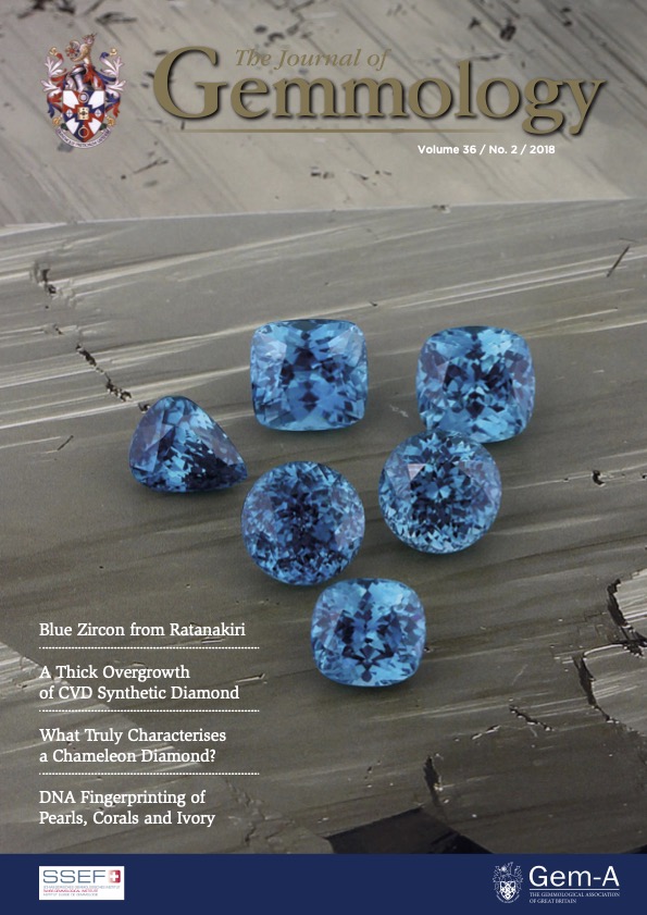
Boehm, E.W. and Laurs, B.M. (2018) Gem Notes: Blue gahnite from Nigeria. Journal of Gemmology, Vol. 38, No. 2, pp. 96–97.
Record No. 6292  |  ISBN/ISSN: 221252  |  Sinkankas/Born No.: NIS/NIB  |  RWHL
Boehm and Laurs describe small, deeply coloured blue gahnites reportedly originating from south-eastern Nigeria near the Cameroon border, a locality distinct from previously studied occurrences near Jemaa in central Nigeria. Gahnite, ZnAl₂O₄, is the zinc-rich member of the spinel group and forms solid solutions with magnesium spinel and hercynite. Earlier studies of Nigerian material attributed its blue colour either to iron-related electronic transitions and intervalence charge transfer or to trace cobalt; the present note does not establish the colour mechanism of the newly examined stones. Samples were brought to the authors’ attention by gem dealer Eric Braunwart at the June 2017 JCK show in Las Vegas, where they were initially presented as blue spinels. The rough ranged from slightly worn, well-formed octahedra to rounded and more strongly waterworn crystals, suggesting possible secondary eluvial or alluvial deposition. A consistent deep blue colour was retained even in faceted stones as small as 1 mm. Reported production consisted of intermittent parcels weighing 10–50 g, mostly yielding melee of 1–3 mm; the largest cut stone reported was a 0.30 ct round brilliant measuring 4 mm. Examination of two faceted specimens weighing 0.17 and 0.19 ct gave refractive-index readings of 1.795, consistent with gahnite. A rough crystal yielded a spot reading of 1.78, and crystal specific gravities ranged from 4.40 to 4.65. These results broadly agree with earlier measurements of Nigerian gahnite. Another faceted stone in the submitted material proved to be sapphire, based on refractive indices of 1.762–1.770 and hexagonal growth zoning, demonstrating that the parcel contained more than one gem species. Microscopic features in the gahnite included rounded colourless crystals resembling apatite, rectangular and flat hexagonal colourless crystals, negative crystals, and clouds of fine particles. The mineral identities of these inclusions were not analytically confirmed. Samples showed a moderate red Chelsea filter reaction. A digital spectroscope revealed strong absorption at 460–465 nm and weaker absorption at 570–590 nm, features comparable to those previously documented in Nigerian gahnite. The authors report strong trade demand despite the material’s small typical size and anticipate that available production will be readily absorbed by the market. The note provides preliminary identification data and commercial context for an uncommon blue gemstone from a reported new Nigerian source.

A gemological description of Ethiopian emeralds
Journal Article
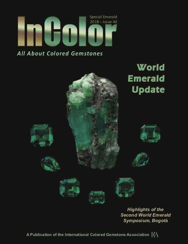
Vertriest, W. and Wongrawang, P. (2018) A gemological description of Ethiopian emeralds. InColor, pp. 73–75.
Record No. 6055  |  Sinkankas/Born No.: NIS/NIB  |  RWHL
In 2016, a new deposit of emeralds was discovered in the rural village of Kenticha and Dermi in the Seba Boru distinct near the town of Shakiso, which is located in the Oromia province in the Southern Ethiopia. GIA had the opportunity to study many samples from this new deposit.

Characteristics of faceted-quality ruby from Longido, Tanzania
Journal Article
Leelawatanasuk, T., Susawee, N. and Bupparenoo, P. (2018) Characteristics of faceted-quality ruby from Longido, Tanzania. Bulletin of Earth Sciences of Thailand, Vol. 9, pp. 1–7.
Record No. 6014  |  Sinkankas/Born No.: NIS/NIB  |  RWHL
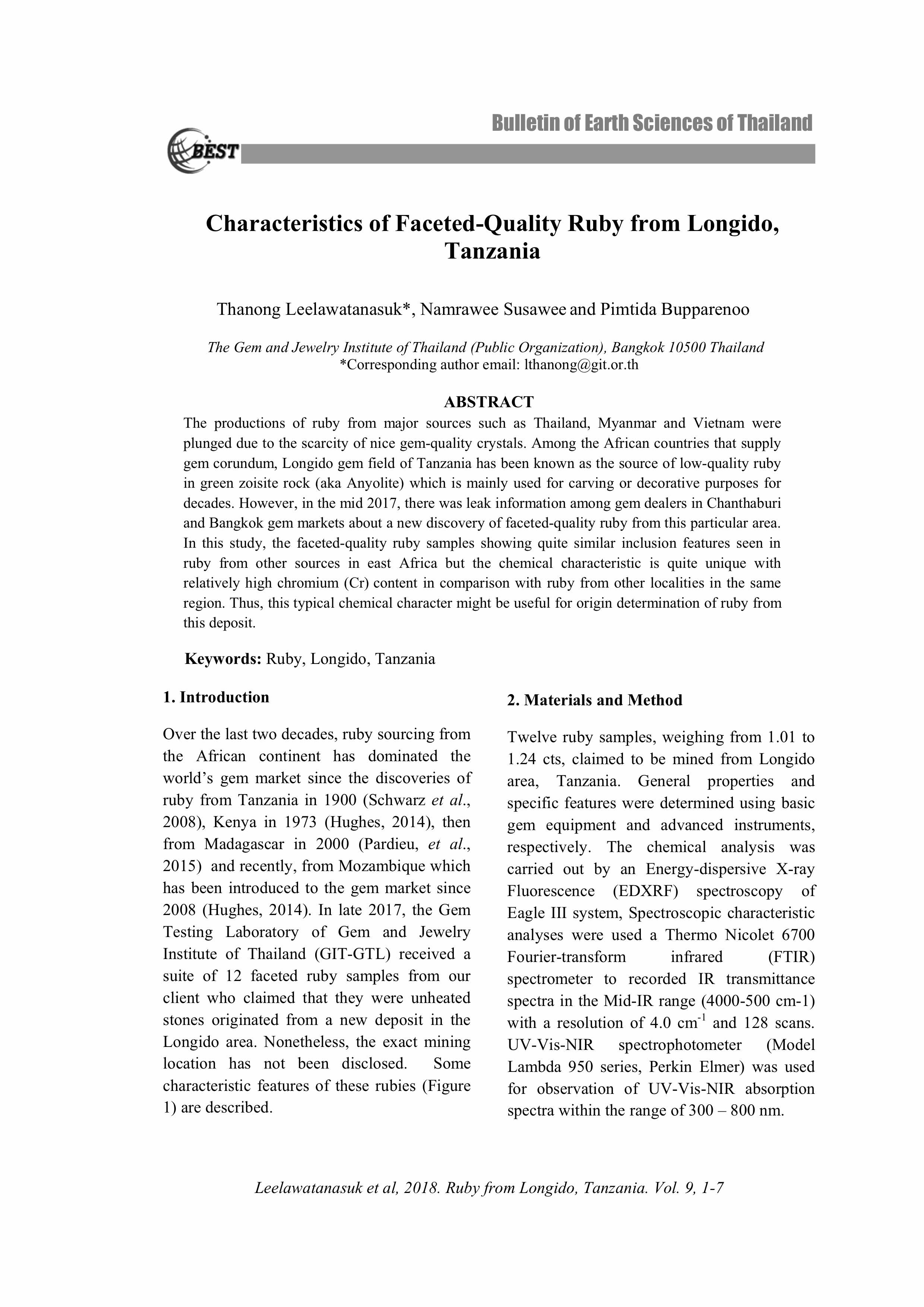
The productions of ruby from major sources such as Thailand, Myanmar and Vietnam were plunged due to the scarcity of nice gem-quality crystals. Among the African countries that supply gem corundum, Longido gem field of Tanzania has been known as the source of low-quality ruby in green zoisite rock (aka Anyolite) which is mainly used for carving or decorative purposes for decades. However, in the mid 2017, there was leak information among gem dealers in Chanthaburi and Bangkok gem markets about a new discovery of faceted-quality ruby from this particular area. In this study, the faceted-quality ruby samples showing quite similar inclusion features seen in ruby from other sources in east Africa but the chemical characteristic is quite unique with relatively high chromium (Cr) content in comparison with ruby from other localities in the same region. Thus, this typical chemical character might be useful for origin determination of ruby from this deposit.

Fluid inclusion study of trapiche and non-trapiche rubies from the Mong Hsu district, Myanmar
Journal Article
Giuliani, G., Dubessy, J., Pignatelli, I. and Schwarz, D. (2018) Fluid inclusion study of trapiche and non-trapiche rubies from the Mong Hsu district, Myanmar. Canadian Mineralogist, Vol. 56, No. 5, pp. 691–703.
Record No. 6012  |  ISBN/ISSN: https://doi.org/10.3749/canmin.1800013  |  Sinkankas/Born No.: NIS/NIB  |  RWHL
The Mong Hsu ruby deposit in Myanmar is a marble-type deposit. It is well-known for attractive crystals with dark violet/blue to black cores and ruby rims, as well as for trapiche rubies. These exceptional rubies have a texture composed of six growth sectors separated by six dendrites running from a central core to the six corners. Fluid inclusions (FI) have been studied in both types of ruby, i.e., trapiche and non-trapiche crystals. Microthermometry combined with Raman spectrometry investigations of primary and secondary FI proved the existence of CO₂–H₂S–COS–S₈–AlO(OH)-bearing fluids with diaspore and native sulfur daughter minerals. The carbonic fluid belongs to the CO₂–H₂S system with CO₂ as a dominant component. Minor COS (1–2 mol.%) and diaspore indicate that H₂O (∼1 mol.%) was present in the paleofluid. The amount of H₂S in FI from trapiche ruby is between 6.9 and 7.4 mol.% for the dendrites and from 7.2 to 15.7 mol.% for the growth sectors. The H₂S content is approximately 10 mol.% in FI from non-trapiche rubies. The presence of dawsonite and previous crushing data carried out on well-cleaned crystals of non-trapiche ruby indicate that an ionic salt melt was present at the time of ruby formation. The non-detection of this melt in trapiche ruby is attributed to the small size of the remnants of molten salts, making it difficult to observe them under the microscope. Following this hypothesis, the FI assemblages in rubies would correspond to the trapping of two immiscible fluids, i.e., a carbonic phase in the CO₂–H₂S–COS–S₈–AlO(OH)-system and molten salts. Such fluid phases are considered to be the product of metamorphism of evaporites during devolatilization of carbonates and thermal-sulfate reduction. The formation of trapiche ruby is discussed in terms of possible variations of the composition of the fluid, temperature, and pressure conditions. The variation in density of the carbonic fluids from the dendrites (0.70 < ρ < 0.78) to the growth sectors (0.56 < ρ < 0.68) records variation of fluid pressure in the metamorphic system. This variation led to episodically local fluid overpressure and hydraulic fracturing in the marble. During such episodes, changes in driving-force conditions allowed for the formation of the trapiche texture in ruby: the development of dendrites and growth sectors occurred under high and low driving-force conditions, respectively. Non-trapiche ruby in veinlets formed concurrently under thermodynamic conditions similar to those registered for the growth sectors in trapiche ruby.

Spinel formation by sulphur-rich saline brines from Mansin (Mogok area, Myanmar)
Web Page
Peretti, A., Mullis, J., Franz, L. and Günther, D. (2018) Spinel formation by sulphur-rich saline brines from Mansin (Mogok area, Myanmar).
Record No. 6001  |  Sinkankas/Born No.: NIS/NIB  |  RWHL
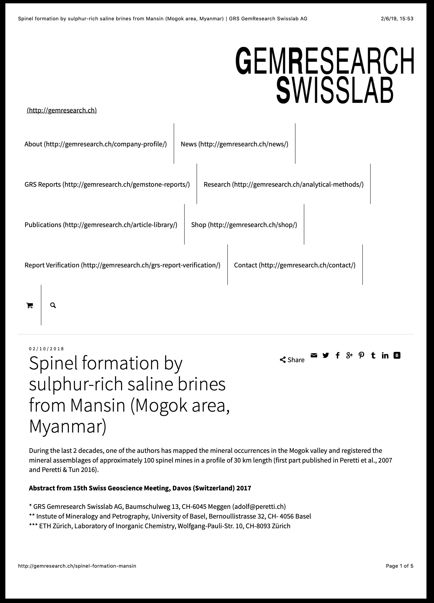
Investigates spinel formation at Mansin through field observations and analysis of fluid and solid inclusions. Spinels occur in impure dolomitic marble with olivine and sulfides. Two inclusion assemblages, interpreted as trapped immiscible fluids, contain hydrogen sulfide, salts and numerous daughter minerals. Raman spectroscopy and microthermometry identify an H₂S-rich phase homogenizing at 101 ± 2°C. The preliminary findings indicate that sulfur-rich, highly saline brines promoted spinel formation.

Sri Lankan sapphire with spinel inclusions
Journal Article
Smith, C.P. (2018) Sri Lankan sapphire with spinel inclusions. Journal of Gemmology, Vol. 36, No. 4, p. 291.
Record No. 5966  |  ISBN/ISSN: 221252  |  Sinkankas/Born No.: NIS/NIB  |  RWHL
Recently, a 5.06 ct yellow sapphire was submitted to the American Gemological Laboratories (AGL) for testing (Figure 18). Standard gemmological analyses distinguished that this was an unheated yellow sapphire originating from Sri Lanka. What caught the eye of this gemmologist were two well-formed octahedral crystals situated under the table of the gem (Figure 19). Raman analyses identified them as spinel. Corundum and spinel often form in similar geological environments and geographical locations. Given this, it is not surprising that spinel (as well as gahno-spinel) has been identified previously in gem-quality corundum from a variety of sources, such as Sri Lanka and Myanmar/Burma (see, e.g., Gübelin and Koivula, 2008). However, it is not as common to see such euhedral colourless crystals of spinel as are present in this yellow sapphire.

G&G Micro-World: Sillimanite in ruby

Journal Article
Schneider, V. and Smith, T. (2018) G&G Micro-World: Sillimanite in ruby. Gems & Gemology, Vol. 54, No. 4, Winter, pp. 448–449.
Record No. 5962  |  ISBN/ISSN: 0016626X  |  Sinkankas/Born No.: NIS/NIB  |  RWHL
A ruby submitted to the Gemological Institute of America (GIA) for origin determination contained unusual sillimanite inclusions identified by laser Raman spectroscopy. Its high iron concentration (7,100–7,410 ppmw), combined with the inclusions, indicated an amphibolite-hosted, non-classic metamorphic origin consistent with East African sources. The dense needle-like inclusions may reduce transparency, explaining their rarity in gem-quality rubies. This case highlights the importance of mineral inclusions and trace-element analysis in determining ruby origin.

Corundum with spinel corona from the Tan Huong-Truc Lau area in norhern Vietnam
Journal Article
Khoi, N.N., Hauzenberger, C., Sutthirat, C., Tuan, D.A., Häger, T. and Nam, N.V. (2018) Corundum with spinel corona from the Tan Huong-Truc Lau area in norhern Vietnam. Gems & Gemology, Vol. 54, No. 4, Winter, pp. 404–417.
Record No. 5961  |  ISBN/ISSN: 0016626X  |  Sinkankas/Born No.: NIS/NIB  |  RWHL
Corundum deposits in the Tan Huong–Truc Lau area of Vietnam are hosted mainly in gneiss and partly in feldspathic pegmatoid and marble. Encountered here in both primary and secondary placer deposits is a considerable quantity of stones with a well-developed spinel corona following the morphology of the corundum core. The genesis of the observed spinel corona can be explained by their reaction textures, chemical composition, and mineral inclusions as well as by applying thermodynamic phase equilibria calculations. The following reactions were found to be responsible for the formation of the spinel corona around corundum in gneiss: biotite + corundum → spinel + K-feldspar + fluid, or garnet+corundum → spinel + sillimanite For corundum-spinel associations in metacarbonate rocks, the relevant reaction is: dolomite + corundum → spinel + calcite + carbon dioxide. Mineralogical and chemical studies have shown that the gem quality of ruby and sapphire grains from Tan Huong–Truc Lau deteriorated substantially after their formation due to this corona texture.

Gem Notes: Blue-to-violet spinels from Mozambique
Journal Article
Borenstein, G. and Sevdermish, M. (2018) Gem Notes: Blue-to-violet spinels from Mozambique. Journal of Gemmology, Vol. 36, No. 2, pp. 104–106.
Record No. 5902  |  ISBN/ISSN: 221252  |  Sinkankas/Born No.: NIS/NIB  |  RWHL
Borenstein, Sevdermish, and Boehm report blue-to-purple spinels attributed to Mozambique, documenting their commercial appearance, measured properties, and internal features while noting that their precise mining localities remain unknown. Rough-stone dealer Farooq Hashmi first encountered several kilograms of purple and blue material in Arusha, Tanzania, in November 2015, with additional offerings during visits in May and June 2016. The rough consisted chiefly of broken pieces weighing less than 3 g. One selected piece was fashioned into a 1.36 ct round brilliant for examination. This slightly violetish blue stone had a refractive index of 1.718 and hydrostatic specific gravity of 3.66. It showed no reaction through the Chelsea filter, no discernible absorption features with a handheld spectroscope, and no inclusions visible under a gemmological microscope. A broader range of reportedly Mozambican spinel appeared at the 2018 Tucson gem shows, encompassing greyish and greenish blue, violet, and purple. Cutting of this material began in early 2017, yielding stones from 2 mm in diameter to 8 ct; approximately 3,000 carats of larger stones had been faceted. Seven examples weighing 1.20–3.83 ct were examined in greater detail. Five ranged from violetish purple to reddish purple, with medium-to-dark tones and moderate-to-vivid saturation, while two were greenish blue to blue, with dark tones and high saturation. Their refractive indices were 1.718 ± 0.001 and hydrostatic specific gravities ranged from 3.58 to 3.62. Microscopic features included fine needles distributed as short silk-like inclusions, elongated networks with an apparently partly dissolved character, and intersecting arrangements producing a spiderweb-like appearance. Fingerprint inclusions and small transparent hexagonal prisms were also observed. The prisms were doubly refractive and resembled apatite, although the note does not report analytical confirmation of that identification. Photographs illustrate the colour range and contrasting inclusion patterns. Differences between the initially examined stone and the later group, particularly in colour appearance and specific gravity, lead the authors to suggest that more than one deposit may be represented. The report expands the documented range of Mozambique spinel beyond previously described pink material, providing preliminary gemmological observations while leaving deposit-level provenance unresolved.

Update on “low-temperature” heat treatment of Mozambican ruby: A focus on inclusions and FTIR spectroscopy
Report
Saeseaw, S., Kongsomart, B., Atikarnsakul, U., Khowpong, C., Vertriest, W. and Soonthorntanikul, W. (2018) Update on “low-temperature” heat treatment of Mozambican ruby: A focus on inclusions and FTIR spectroscopy. GIA News from Research, Bangkok: Gemological Institute of America, 25-Apr, 45 pp.
Record No. 5868  |  Sinkankas/Born No.: NIS/NIB  |  RWHL
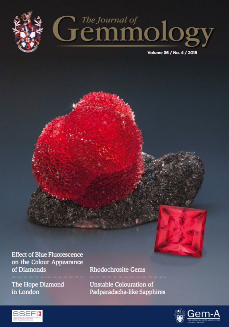
Gemstones have been treated for as long as they have been used by humans. The supply of untreated high-quality gems is so incredibly small that people have always tried to find ways to optimize the other material found in the mines. Treatments come in many different forms and degrees of quality improvement. Dyeing to create color, oiling to hide fractures, heat treatment to increase clarity and optimize color, irradiation to modify color, and impregnation for better stability are just a few examples (Nassau, 1981). In the case of corundum, heat treatment is the most common form. By applying the correct heating process, color can be introduced, strengthened, reduced, or removed. These variations are determined by an interplay between the gemstones, temperature profile during treatment, duration of heating, heating atmosphere (reducing or oxidizing), and the presence of added chemicals (Hughes, 2016; Emmett et al., 2003, 1993). Recent developments have caused drastic changes in heat treatment applications. Since 2009, Mozambique has been an important source of rubies in the world trade. It was quickly discovered that these stones reacted well to a form of heat treatment that used lower temperatures. This modern treatment at low temperatures is distinguished from the classic high-temperature methods by comparing their effect on the minute particles in the stones. These second-phase microcrystals are most commonly rutile (Ti-oxide) that takes the form of fine clouds or silk. During low-temperature treatment, these inclusions are not dissolved in the corundum lattice. This is clearly the case for high-temperature treatment, where silk is obviously altered or even completely diffused. Since heat treatment depends on more than the temperature alone, it is very hard to state a clear temperature boundary between low-temperature and high-temperature treatment. The most important factors to consider in this case are temperature and time. The speed of the process increases considerably with higher temperatures, but longer heating periods at lower temperatures might yield similar results. A rough guideline for the temperature boundary is somewhere between 1200 and 1350°C. Preliminary studies on low-temperature treatment have been published (Pardieu, Saeseaw et al., 2015; Sripoonjan, Wanthanachaisaeng et al., 2016). Both of these publications mention subtle alterations, notably in stained fractures and silk platelets. Spectroscopy showed promising results, but the results were inconclusive due to the small sample sizes. Because heat treatment processes are constantly evolving, updated data needs to be collected regularly. The objective of this project is to observe changes in Mozambican rubies during known treatment conditions (temperature and duration). The focus is on inclusions and FTIR spectra after heat treatment.

Peculiar trapiche-like pattern in a gold-sheen sapphire
Journal Article
Bui, T.N. (2018) Peculiar trapiche-like pattern in a gold-sheen sapphire. Journal of Gemmology, Vol. 36, No. 4, pp. 289–291.
Record No. 4099  |  ISBN/ISSN: 221252  |  Sinkankas/Born No.: NIS/NIB  |  RWHL
Describes a gold-sheen sapphire from Kenya.

G&G Micro-World: Zircon cluster in Ethiopian sapphire
Journal Article
Khowpong, C. (2018) G&G Micro-World: Zircon cluster in Ethiopian sapphire. Gems & Gemology, Vol. 54, No. 4, Winter, pp. 450–451.
Record No. 4085  |  ISBN/ISSN: 0016626X  |  Sinkankas/Born No.: NIS/NIB  |  RWHL

G&G Lab Notes: Large pargasite inclusion in Kashmir sapphire
Journal Article
Muyal, J. (2018) G&G Lab Notes: Large pargasite inclusion in Kashmir sapphire. Gems & Gemology, Vol. 54, No. 4, Winter, pp. 435–436.
Record No. 4083  |  ISBN/ISSN: 0016626X  |  Sinkankas/Born No.: NIS/NIB  |  RWHL

Chemistry and gemmological features of spinels from Sri Lanka and Vietnam
Conference Paper
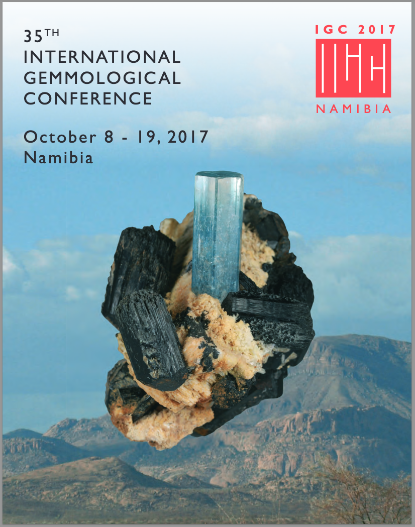
Kleišmantas, A., Zoysa, G.E. and Liu, S. (2017) Chemistry and gemmological features of spinels from Sri Lanka and Vietnam. 35th International Gemmological Conference Proceedings, 8–19 October, Windhoek, Namibia pp. 148–150.
Record No. 29505  |  Sinkankas/Born No.: NIS/NIB  |  RWHL
A study on the chemistry and gemmological features of spinels from Ratnapura in Sri Lanka and Luc Yen in Vietnam using scanning electron microscopy. The research identifies chromophore oxides responsible for coloration, noting that V³⁺ determines yellow and brown hues, Cr³⁺ drives red and pink tones, and Fe²⁺ with low Co²⁺ produces purple or violet shades. Mineral inclusions were cataloged, with Vietnamese spinels containing dolomite, calcite, titanite, rutile, zircon, graphite, apatite, pyrrhotite, pentlandite, violarite, clinochlore, phlogopite, magnesite and chondrodite, while Sri Lankan samples featured allanite, paragonite, zoisite, K-feldspar, plagioclase, florencite, biotite, pyrite and diopside. The authors conclude that vanadium oxide content above 0.4 wt.% is critical for orange and brown coloration in spinels, complementing existing models that primarily rely on chromium, iron and cobalt.

Microscopy as a form of art

Journal Article
Avramouli, A., Gonidi, M. and Lambropoulou, M. (2017) Microscopy as a form of art. Technoetic Arts, Vol. 15, No. 2, pp. 195–202.
Record No. 29391  |  ISBN/ISSN: 1758-9533  |  DOI: https://doi.org/10.1386/tear.15.2.195_1  |  Sinkankas/Born No.: NIS/NIB  |  RWHL
Abstract Art and Science are usually considered fields of different perceptions and approaches with very little overlap, but when combined, sometimes something unusually beautiful and unexpected can arise. Currently, a major effort is being made to promote the interaction between artists and scientists as demonstrated by some of the most recent grant programmes by organizations such as the United Kingdom’s Engineering and Physical Sciences Research Council (EPSRC). Occasionally, artists are approaching science with slightly different insights than those expected, but to date, there is a gap that is rarely violated despite the extraordinary ideas that may arise from such collaborations. There can be no doubt that Nature is the greatest inspiration for art and beauty. In many ways, science attempts to reveal the rules and the beauty of nature. Microscopy, as a technique in scientific research, plays a key role in revealing the beauty of nature, a trend that has led some to regard it as a form of art. But can this claim be justified? For the uninitiated, microscopy could provide a high-resolution image of nature’s creations, revealing at the same time its inherent beauty. The cells – human or non-human – form images of the same beauty as those represented in paintings. Preparations observed under the microscope contain a wealth of information. The aim of the researcher is to correlate the cellular structure with the functioning of the organism and ultimately with the miracle of life. The aesthetic quality of the images has led many scientists to stand in their artistic nature. But can we say that microscopy, which is essentially a way of acquiring and analyzing scientific data, is a form of art? If art is the creation of works by people of extraordinary talent, then microscopy might be an art form and a technique that seeks the functional disclosure of complex admirers of cellular structures. However, unlike music or painting, microscopy is certainly not a form of art, with the traditional concept of creativity. The question therefore arises as to whether the aesthetic nature of the images revealed by a microscope can be considered as art. This is a question that is the starting point of this article.

The linkage between garnets found in India at the Arikamedu archaeological site and their source at the Garibpet deposit
Journal Article
Schmetzer, K., Gilg, H.A., Schüssler, U., Panjikar, J., Calligaro, T. and Périn, P. (2017) The linkage between garnets found in India at the Arikamedu archaeological site and their source at the Garibpet deposit. Journal of Gemmology, Vol. 35, No. 7, pp. 598–627.
Record No. 17211  |  ISBN/ISSN: 221252  |  DOI: http://dx.doi.org/10.15506/JoG.2017.35.7.598  |  Sinkankas/Born No.: NIS/NIB  |  RWHL
The archaeological site of Arikamedu, located in Tamil Nadu State on the east coast of India, was for many centuries the center of a significant bead-producing industry. Beads were made from both glass and stone, including garnet, though the source of the garnet rough material had not been confirmed. To investigate this, garnet beads found at Arikamedu were compared with rough material from the Garibpet deposit, located approximately 640 km away in Telangana State, east of Hyderabad. Samples from both locations showed substantial similarities in average composition, trace-element contents, chemical zoning of major and minor elements, inclusion assemblages, and zoning of inclusions between the rims and cores of the crystals. Chemically, the garnets were almandine-rich (averaging 81.0% almandine, 11.5% pyrope, 3.3% spessartine, and 1.5% grossular), with clear zoning for manganese (Mn) and magnesium (Mg), as well as trace elements such as yttrium (Y), phosphorus (P), and zinc (Zn). The most distinctive aspect of the inclusion pattern was the presence of sillimanite fibers concentrated in a zone between the inclusion-rich core and the inclusion-poor rim. Combining microscopic observations, inclusion identification, and chemical analyses, it was determined that the rough material used to produce garnet beads at Arikamedu historically originated from the Garibpet deposit. Additionally, the results suggest that current classification systems for historical garnets need further refinement.

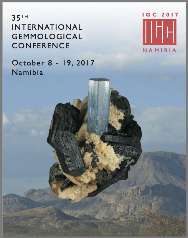
Study and investigation of spinel from Kermunda in Odisha, India
Conference Paper
Panjikar, J. and Panjikar, A. (2017) Study and investigation of spinel from Kermunda in Odisha, India. International Gemmological Conference, 8–19 October, Windhoek, Namibia: International Gemmological Conference, pp. 62–66.
Record No. 7804  |  Sinkankas/Born No.: NIS/NIB  |  RWHL
Spinel found in the Kermunda village (19.9671° N, 82.7913° E) located in Golamunda Tehsil of Kalahandi district of Odisha state of India were investigated. Odisha was formerly known as Orissa state and has been known for gemstone deposits. In Kermunda, in Kalahandi (Fig1) the area constitutes a part of the Precambrian khondalite-charnockite-granite gneiss terrain of Eastern Ghats having thick soil horizon. It is common to find spinel along with corundum and other minerals in this soil. Locals collect red coloured broken spinel crystals, calling them “naram” meaning low grade “ruby”, to make beads. Gemmological properties, values for refractive indices, specific gravity, as well as FTIR and UV-Vis data have been determined for the Kermunda spinel. Semi-quantitative analyses carried out using EDXRF show considerable presence of titanium and iron oxides in the spinel. Inclusion studies by Raman microspectroscopy identified mineral inclusions as apatite, ilmenite, rutile, spinel and zircon. It is the first time that högbomite has been reported as an inclusion in gem-quality Indian spinel.

The genesis and SHRIMP U–Pb zircon dating of the Pishan brown nephrite-bearing Mg–skarn deposit in Xinjiang
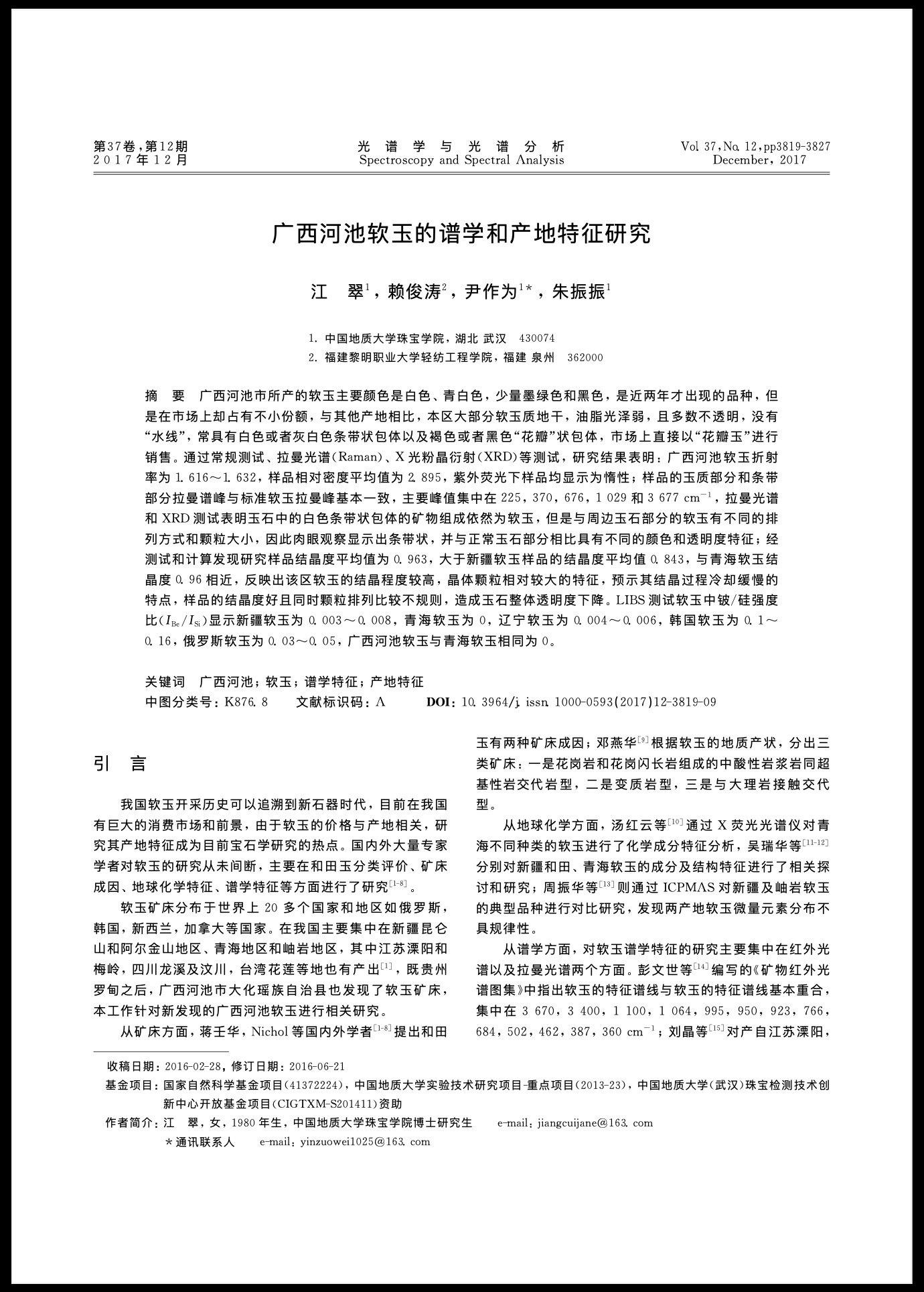
新疆皮山镁质矽卡岩矿床(含糖玉)成因及锆石 SHRIMP U-Pb 定年
Journal Article
Liu, X., Liu, Y., Li, Z., Maituohuti, A., Tian, G. and Guo, D. (2017) The genesis and SHRIMP U–Pb zircon dating of the Pishan brown nephrite-bearing Mg–skarn deposit in Xinjiang. [新疆皮山镁质矽卡岩矿床(含糖玉)成因及锆石 SHRIMP U-Pb 定年] Acta Petrologica et Mineralogica [岩石矿物学杂志], Vol. 36, No. 2, March, pp. 259–273.
Record No. 7251  |  Sinkankas/Born No.: NIS/NIB  |  Language: in Chinese with English abstract  |  RWHL
The Hetian nephrite belt in Xinjiang, China, is the world’s longest nephrite belt, stretching approximately 1,300 km, and now includes a recently identified type of brown nephrite found in Pishan County. This nephrite occurs in a skarn zone between Changchengian dolomitic marble and Late Hercynian granodiorite, formed by metasomatic processes at the contact. Microscopically, the brown nephrite consists mainly of fibrous tremolite with minor associated minerals, and its brown coloration is structurally disseminated, appearing in fissures as veinlets or leaf-like forms. EMPA and XRD analyses show it is composed of tremolite along with illite, Mg-olivine, diopside, chromite, and other accessory phases. Tremolite developed through metasomatism with dolomitic marble and by replacing diopside. Whole-rock geochemistry reveals strong negative Eu anomalies (δEu = 0.09–0.28), depleted LREEs, flat HREEs, and low overall REE concentrations, patterns closely resembling those of the host dolomitic marble, supporting a genetic link. Stable isotope data from ore-forming fluids at 330 °C (δD = –81.0‰ to –84.0‰; δ¹⁸O = 3.16‰ to 5.48‰) align with those of marble-type nephrite and differ markedly from serpentine-type nephrite, suggesting that the fluid was a mixture of magmatic and meteoric water with CO₂ from decarbonated marble. Ar–Ar dating of phlogopite indicates a formation age of 285.23 ± 1.57 Ma, while SHRIMP U–Pb dating of zircon inclusions gives an older age of 456 ± 7 Ma, suggesting that the nephrite formed after the incorporation of pre-existing zircon, supporting a Late Hercynian metasomatic origin.

Research on spectroscopy and locality characteristics of nephrite jade from Hechi, in Guangxi
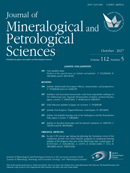
广西河池软玉的谱学和产地特征研究
Journal Article
Jiang, C., Lai, J., Yin, Z. and Zhu, Z. (2017) Research on spectroscopy and locality characteristics of nephrite jade from Hechi, in Guangxi. [广西河池软玉的谱学和产地特征研究] Spectroscopy and Spectral Analysis [光谱学与光谱分析], Vol. 37, No. 12, December, pp. 3819–3827.
Record No. 6976  |  Sinkankas/Born No.: NIS/NIB  |  Language: in Chinese with English abstract  |  RWHL
The nephrite, which has been found in Hechi, Guangxi in the past couple of years, in various colors like white, greenish-white, rarely dark green and black, is gaining a growing popularity. Compared with those located in other places, the Hechi nephrite presents a greasy luster, which has a dry and mostly opaque texture without “water lines.” The inclusions, of which are band patterns in white or grayish-white colors and dendritic patterns in brownish-yellow and dark colors. That’s why it is named “dendritic jade” in the market. The samples were tested by standard gemological methods, Raman spectroscopy, X-ray diffraction (XRD), and other techniques. The research results show that the RI of them is from 1.616 to 1.632, an average hydrostatic S.G. 2.895, inert under UV. The Raman spectra of jade part and white band are consistent with the standard Raman spectrum of nephrites. The main peaks are mainly 225, 370, 676, 1,029, and 3,677 cm⁻¹. Despite the results of the Raman and XRD showing that the white bands are nephrites, the band parts show different color and transparency with naked eyes because of different display patterns and sizes of the grains in the bands. Through test and calculation, the degree of crystallinity of research samples is 0.963 on average which is bigger than that of Xinjiang 0.843 on average and similar to that of Qinghai 0.96 on average. This means nephrites in this era crystallized well so as to make the mineral grains bigger, which indicates the characteristics of slow cooling crystallization process. Because the crystallinity of the samples is good and grains are more irregular at the same time, the overall transparency of jade decreased. The value IBe/ISi of LIBS test on nephrites located in Xinjiang is 0.003–0.008, Qinghai 0, Liaoning 0.004–0.006, Korea 0.1–0.16, Russia 0.03–0.05, while that of Hechi in Guangxi is 0, similar to that of Qinghai.

Chemical composition of fluid inclusions in the Yorii jadeite–quartz rocks from the Kanto Mountains, Japan

Journal Article
Fukuyama, M., Kawamoto, T. and Ogasawara, M. (2017) Chemical composition of fluid inclusions in the Yorii jadeite–quartz rocks from the Kanto Mountains, Japan. Journal of Mineralogical and Petrological Sciences, Vol. 112, No. 5, pp. 281–290.
Record No. 6692  |  DOI: https://dx.doi.org/10.2465/jmps.170331  |  Sinkankas/Born No.: NIS/NIB  |  RWHL
Jadeite–quartz rocks from the Yorii area in the Kanto Mountains of central Japan occur as tectonic blocks within a serpentinite mélange. Primary two-phase (liquid + vapor) aqueous fluid inclusions have been identified in jadeite, quartz, albite, and zircon. The fluids trapped in jadeite and quartz consist of H₂O, with or without CH₄, and display a broad range of salinities—4.7 ± 1.1 wt% NaCl equivalent in jadeite, and 3.4 ± 0.7 and 13.6 ± 2.0 wt% NaCl equivalent in quartz. This study analyzes the chemical composition of fluid inclusions in quartz from jadeite–quartz rocks to elucidate the nature of fluids involved in rock formation within a subduction zone. Trace element analysis using LA–ICP–MS reveals that these fluid inclusions are enriched in large-ion lithophile elements (LILE), lithium (Li), boron (B), high field strength elements (HFSE), and transition metals. The fluids show LILE-enriched and relatively HFSE-depleted patterns, resembling the signature of HFSE-depleted fluids released from antigorite during subduction, although the absolute concentrations differ. These signatures may represent common chemical characteristics of subduction-zone fluids. Two possible scenarios are proposed for the chemical evolution of these fluids following the formation of jadeite–quartz rocks: (1) HFSE may be sequestered into the jadeite–quartz assemblage, resulting in evolved fluids that are relatively depleted in HFSE and enriched in LILE before migrating into the mantle wedge; (2) the fluids may remain in chemical equilibrium with the jadeite–quartz rocks, retaining similar compositions to the inclusions observed. In either case, the metasomatic formation of jadeite–quartz rocks produces residual fluids enriched in LILE and depleted in HFSE. This geochemical behavior mirrors that of arc magmas, which are typically LILE-enriched and HFSE-depleted. The observed elemental signatures in jadeite–quartz fluid inclusions thus provide a mechanism to explain the characteristic geochemistry of magmas in subduction zones.

Exploring the Literature of Gemstone Inclusions

The literature of gemstone inclusions covers identification, origin determination, treatments, synthesis, mineral inclusions, fluid inclusions and other internal features of natural and synthetic gems.

For a curated selection of important sources, read Inclusions in Gemstones — Hyperion Literature Sources.

About Four Treasures

Four Treasures is Lotus Gemology's freely accessible gemological reference database. Use the full database to search by author, title or keyword and explore literature on gemology, mineralogy, jewelry and related fields. Where available, hyperlinks on titles may lead directly to the full publication.

Search the Complete Four Treasures Database