
Gemstone Inclusions Bibliography

Gemstone Inclusions Bibliography

Explore books, articles and historical sources on inclusions in gemstones in Four Treasures, the Lotus Gemology reference database. This bibliography brings together matching records from the collection, with publication details, abstracts and cover images where available.

Gemstone Inclusion References

The list below matches the terms inclusion or inclusions in the database's searchable fields. Some references discuss inclusions as part of a broader subject.
521–540 of 1,247 matching references

The genetic linkage between the Yuanjiang marble-hosted ruby deposit and Cenozoic tectonic evolution of the Ailao Shan-Red River shear zone (Southwest China)
Journal Article
Yang, T., Sun, X., Shi, G., Li, D. and Zhou, H. (2019) The genetic linkage between the Yuanjiang marble-hosted ruby deposit and Cenozoic tectonic evolution of the Ailao Shan-Red River shear zone (Southwest China). Journal of Asian Earth Sciences, Vol. 177,, 2019/06/15/ pp. 38–47.
Record No. 7376  |  ISBN/ISSN: 13679120  |  DOI: https://doi.org/10.1016/j.jseaes.2019.03.010  |  Sinkankas/Born No.: NIS/NIB  |  RWHL
The Yuanjiang marble-hosted ruby deposit, located in the central segment of the Ailao Shan–Red River (ASRR) shear zone, is the only economically viable ruby deposit in China. Hosted within Paleoproterozoic sillimanite gneisses, the ore bodies are found in metamorphosed platform carbonates, where rubies are unevenly distributed in calcite-rich marble layers and primarily associated with phlogopite, scapolite, graphite, and pyrite. Inclusions within the rubies include zircon, rutile, apatite, and calcite. The genesis of the deposit and its relationship to the ASRR shear zone remain incompletely understood. This study constrains the timing of ruby formation using in-situ LA-ICP-MS U–Pb dating of zircon inclusions in ruby and ^40Ar/^39Ar dating of syngenetic phlogopite from ruby-bearing marble. The zircon inclusions yield an age of 36.2 ± 1.1 Ma (MSWD = 1.4, 2σ, n = 4), which falls within the timeframe of crustal thickening and subduction-collision events in the region. Because the ruby deposits are not spatially confined to the shear zone, the ruby formation is interpreted as having been triggered by local continental subduction-collision rather than large-scale left-lateral shearing. The ^40Ar/^39Ar dating of phlogopite (22.2–22.5 Ma) provides a minimum estimate for the ore-forming age, given that the closure temperature of this system is lower than the temperature of ruby formation. These younger phlogopite ages, compared to those from deposits outside the shear zone (e.g., Luc Yen), suggest a slower cooling rate for deposits within the shear zone, likely due to continued thermal input from shearing activity. Consequently, while large-scale shearing was not the primary cause of ruby formation, it likely played a key role in transporting marble-hosted ruby deposits within the ASRR zone toward the subsurface, contributing to their preservation and exposure.

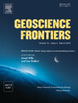
Mineralogical study on nephrite from No.7 deposit, QieMo, Xinjiang
新疆且末 7号矿和 田玉 的矿物学特征
Thesis
Zhang, Y. (2019) Mineralogical study on nephrite from No.7 deposit, QieMo, Xinjiang. [新疆且末 7号矿和 田玉 的矿物学特征]., Beijing, China: China University of Geosciences, 87 pp.
Record No. 7313  |  Sinkankas/Born No.: NIS/NIB  |  Language: in Chinese with English abstract  |  RWHL
The No. 7 nephrite deposit is located in Qiemo, Xinjiang, northwest China, within the Kunlun–Altyn nephrite belt along the southern margin of the Tarim Basin. Field observations and hand specimen analysis reveal distinct mineralization zoning: a magnesian marble belt, a serpentine-altered marble and gray-white nephrite contact belt, a diopside-altered marble belt, a gray nephrite belt, and a granite intrusion belt. The gray, gray-white, and white nephrite is relatively pure, composed mainly of tremolite, with minimal inclusions aside from scattered apatite, chlorite, and mica. It is inferred that these nephrites formed from hot hydrothermal fluids during a short-term rapid cooling phase of metasomatism. Major element analysis suggests that color zoning is related to Fe and Mn content, with increasing concentrations corresponding to a shift from white to green. Trace and rare earth element levels are low overall, with enrichment in LREE and relative depletion in Nb, Sr, and Hf, while U, La, P, and Ti are enriched. A negative Eu anomaly (0.18–0.51) suggests plagioclase separation during crystallization. Polarizing microscope and BSE imagery show high-quality nephrite with a feather-like micro-interlaced texture. Textural replacement relationships—calcite by diopside and tremolite, and later fine-grained tremolite replacing coarse-grained diopside and tremolite—indicate the nephrite formed through multiple stages of magmatic hydrothermal metasomatism.

Mineral association and graphite inclusions in nephrite jade from Liaoning, northeast China: Implications for metamorphic conditions and ore genesis
Journal Article
Zhang, C., Yu, X. and Jiang, T. (2019) Mineral association and graphite inclusions in nephrite jade from Liaoning, northeast China: Implications for metamorphic conditions and ore genesis. Geoscience Frontiers, Vol. 10, No. 2, March, pp. 425–437.
Record No. 6229  |  Sinkankas/Born No.: NIS/NIB  |  RWHL
The Liaoning Province in the northeastern part of the North China Craton (NCC) hosts several tremolite jade (nephrite) deposits. Here we investigate the Sangpiyu tremolite jade deposit where the relationship between abundant graphite inclusions within the jade remains enigmatic. We employ petrography, electron probe microanalysis, X-ray-diffraction, and Raman spectroscopy to characterize the tremolite jade and its inclusion minerals. The Sangpiyu jade is predominately composed of tremolite with minor calcite, dolomite, serpentine, titanite, zoisite, allanite, chlorite, apatite, chromite and graphite. Raman spectroscopy of graphite inclusions shows that the D1/G intensity ratio ranges from 0.78 to 0.88 in deep green samples and from 0.05 to 0.23 in dark green samples. The ranges of D1/(D1 + G) integral area ratio for these types are from 0.0548 to 0.3037 and 0.5528 to 0.7355 respectively. The formation temperature of graphite inclusions in the dark green tremolite jade is computed as 549.8°C, whereas that for the deep green sample is about 343.2°C. Our results suggest that the jade formation occurred in a multi-stage process through the action of hydrothermal fluids and metamorphism possibly in a subduction-related setting at moderate to high temperatures.

Gemmological analysis of a Roman sapphire intaglio and Its possible origin
Journal Article
Krzemnicki, M.S., Butini, F., Butini, E. and Carolis, E., de (2019) Gemmological analysis of a Roman sapphire intaglio and Its possible origin. Journal of Gemmology, Vol. 36, No. 8, pp. 710–724.
Record No. 6123  |  ISBN/ISSN: 221252  |  DOI: https://doi.org/10.15506/JoG.2019.36.8.710  |  Sinkankas/Born No.: NIS/NIB  |  RWHL
The gemmological analysis of a Roman intaglio engraved with a hippocamp (winged ‘sea-horse’) reveals that it was carved from a sapphire of basaltic origin. Its bluish grey appearance is due to Rayleigh scattering by sub-microscopic inclusions and is not related to an intervalence charge transfer process. In light of historically documented extensive trade relations between ancient Rome and Ethiopia (the kingdom of Aksum), we hypothesise that the recently documented basalt-related sapphire deposits in northern Ethiopia are a possible source of raw material for this Roman intaglio (as well as other basaltic sapphires used in Roman times), in addition to previously held views that ancient sapphires originate from deposits in Sri Lanka, France and perhaps South East Asia. This rare opportunity to characterise in detail one of the very few engraved sapphires from the Roman period permits a better understanding of gem materials used in classical antiquity.

MicroWorld: Sapphire inclusion with rutile “silk” in a Burmese star sapphire
Journal Article
Jia, X. and Sit, M.M. (2019) MicroWorld: Sapphire inclusion with rutile “silk” in a Burmese star sapphire. Gems & Gemology, Vol. 55, No. 1, Spring, pp. 114–115.
Record No. 5999  |  ISBN/ISSN: 0016626X  |  Sinkankas/Born No.: NIS/NIB  |  RWHL
GIA’s Hong Kong laboratory recently examined a remarkable sapphire inclusion inside a Burmese star sapphire. Possessing the same refractive index as its host, the sapphire guest displays extremely low relief and is nearly invisible under transmitted light. Nevertheless, its outline became distinctive under cross-polarized illumination, showing strong interference colors (figure 11, left). Closer examination using fiber-optic illumination revealed a group of oriented rutile “silk” in this sapphire inclusion (figure 11, right). Although exsolved rutile needles are a common inclusion in Burmese sapphire, it is interesting to see two sets of dense silk exsolved from both the star sapphire host and a protogenetic sapphire inclusion, each aligned with the basal plane of its own host.

Diversity in ruby geochemistry and Its inclusions: Intra- and inter-continental comparisons from Myanmar and eastern Australia
Journal Article
Sutherland, F.L., Khin Zaw, Meffre, S., Thompson, J., Goemann, K., Kyaw Thu, Than Than Nu, Zin, M.M. and Harris, S.J. (2019) Diversity in ruby geochemistry and Its inclusions: Intra- and inter-continental comparisons from Myanmar and eastern Australia. Minerals, Vol. 9, No. 1, 28 pp..
Record No. 5978  |  Sinkankas/Born No.: NIS/NIB  |  RWHL
Ruby in diverse geological settings leaves petrogenetic clues, in its zoning, inclusions, trace elements and oxygen isotope values. Rock-hosted and isolated crystals are compared from Myanmar, SE Asia, and New South Wales, East Australia. Myanmar ruby typifies metasomatized and metamorphic settings, while East Australian ruby xenocrysts are derived from basalts that tapped underlying fold belts. The respective suites include homogeneous ruby; bi-colored inner (violet blue) and outer (red) zoned ruby; ruby-sapphirine-spinel composites; pink to red grains and multi-zoned crystals of red-pink-white-violet (core to rim). Ruby ages were determined by using U-Pb isotopes in titanite inclusions (Thurein Taung; 32.4 Ma) and zircon inclusions (Mong Hsu; 23.9 Ma) and basalt dating in NSW, >60–40 Ma. Trace element oxide plots suggest marble sources for Thurein Taung and Mong Hsu ruby and ultramafic-mafic sources for Mong Hsu (dark cores). NSW rubies suggest metasomatic (Barrington Tops), ultramafic to mafic (Macquarie River) and metasomatic-magmatic (New England) sources. A previous study showed that Cr/Ga vs. Fe/(V + Ti) plots separate Mong Hsu ruby from other ruby fields, but did not test Mogok ruby. Thurein Taung ruby, tested here, plotted separately to Mong Hsu ruby. A Fe-Ga/Mg diagram splits ruby suites into various fields (Ga/Mg < 3), except for magmatic input into rare Mogok and Australian ruby (Ga/Mg > 6). The diverse results emphasize ruby’s potential for geographic typing.

G&G Micro-World: Pyrope-almandine garnet in sapphire host
Journal Article
Ng-Pooresatien, N. (2019) G&G Micro-World: Pyrope-almandine garnet in sapphire host. Gems & Gemology, Vol. 55, No. 2, Summer, p. 263.
Record No. 4159  |  ISBN/ISSN: 0016626X  |  Sinkankas/Born No.: NIS/NIB  |  RWHL
In gemology, garnet occurs in a wide variety of colors, including red, orange, yellow, and green. Garnet is also found as inclusions in diamond, quartz, topaz, aquamarine, and zircon (E.J. Gübelin and J.I. Koivula, Photoatlas of Inclusions in Gemstones, Vol. 1, ABC Edition, Zurich, 1986, pp. 158–160).GIA’s Bangkok laboratory recently received a 3.97 ct transparent greenish blue sapphire for a colored stone identification report. Standard gemological testing and advanced analysis revealed properties consistent with natural sapphire. Interestingly, microscopic examination with a combination of darkfield and oblique fiber-optic illumination revealed a well-formed brownish orange crystal cluster with reflective needles (figure 7). The crystal was singly refractive, and Raman spectrometry matched it with pyrope-almandine garnet. Sapphires with garnet inclusions are rarely seen in laboratory examination, although they are occasionally found in samples from Tanzania and the state of Montana (E.J. Gübelin and J.I. Koivula, Photoatlas of Inclusions in Gemstones, Vol. 3, Opinio Publishers, Basel, Switzerland, 2008, pp. 228–242).

G&G Micro-World: Euhedral phantom sapphire in sapphire
Journal Article
LeCroy, B. (2019) G&G Micro-World: Euhedral phantom sapphire in sapphire. Gems & Gemology, Vol. 55, No. 2, Summer, pp. 263–264.
Record No. 4158  |  ISBN/ISSN: 0016626X  |  Sinkankas/Born No.: NIS/NIB  |  RWHL
A phantom sapphire crystal within a host sapphire crystal was recently examined at GIA’s Carlsbad laboratory. The inclusion showed euhedral morphology as one half of a partially tapered bipyramid. The visible boundaries of the included crystal were composed of unaltered fingerprints with geometric patterns and fine linear rows. The crystal’s surface also showed moderate-order birefringence colors; these represented the shared interface between the host’s crystal lattice and the inclusion’s crystal lattice. External local fingerprints also terminated around the crystal’s end point. Negative crystals within the inclusion showed tabular hexagonal habit. When the host sapphire was exposed to temperatures below the freezing point of water, a CO₂ bubble was seen within the largest negative crystal. A sapphire with this inclusion combination is rarely seen.

Gem News International: Sapphires from Colombia
Journal Article
Rizzo, J. (2019) Gem News International: Sapphires from Colombia. Gems & Gemology, Vol. 55, No. 1, Spring, pp. 155–156.
Record No. 4149  |  ISBN/ISSN: 0016626X  |  Sinkankas/Born No.: NIS/NIB  |  RWHL
The article examines five unheated sapphires from the Mercaderes deposit in southern Colombia, including pink, blue, and colour-change varieties. Gemological testing confirmed their identity as corundum, while LA-ICP-MS revealed high iron concentrations suggesting a magmatic origin. Infrared spectroscopy showed no evidence of treatment, and Raman spectroscopy identified unaltered zircon and rutile inclusions. These findings supported the conclusion that the sapphires were unheated and provided valuable data for gemstone origin determination.

MicroWorld: Star-like growth in natural yellow sapphire
Journal Article
Atikarnsakul, U. (2019) MicroWorld: Star-like growth in natural yellow sapphire. Gems & Gemology, Vol. 55, No. 1, Spring, p. 115.
Record No. 4148  |  ISBN/ISSN: 0016626X  |  Sinkankas/Born No.: NIS/NIB  |  RWHL
GIA’s Hong Kong laboratory recently examined a remarkable sapphire inclusion inside a Burmese star sapphire. Possessing the same refractive index as its host, the sapphire guest displays extremely low relief and is nearly invisible under transmitted light. Nevertheless, its outline became distinctive under cross-polarized illumination, showing strong interference colors (figure 11, left). Closer examination using fiber-optic illumination revealed a group of oriented rutile “silk” in this sapphire inclusion (figure 11, right). Although exsolved rutile needles are a common inclusion in Burmese sapphire, it is interesting to see two sets of dense silk exsolved from both the star sapphire host and a protogenetic sapphire inclusion, each aligned with the basal plane of its own host.

MicroWorld: Trapiche-like ruby from the Batakundi mine, Kashmir
Journal Article
Wongrawang, P. (2019) MicroWorld: Trapiche-like ruby from the Batakundi mine, Kashmir. Gems & Gemology, Vol. 55, No. 1, Spring, p. 114.
Record No. 4147  |  ISBN/ISSN: 0016626X  |  Sinkankas/Born No.: NIS/NIB  |  RWHL
Recently, the author examined a 5.26 ct faceted transparent oval ruby that showed a well-formed fixed six-rayed pattern under the table of the stone. Chemical analysis and gemological observation of internal features determined the origin of this stone to be the Batakundi mining area of Kashmir. The six rays were oriented perpendicular to the sides of the hexagonal growth structure (figure 10). Each arm consisted of numerous minute particles mixed with needle-like inclusions and small thin films. The center of the star contained a group of colorless and black mineral inclusions that were identified by Raman spectroscopy as feldspar and graphite. Near the core, two black crystals with high luster were identified by Raman spectroscopy as rutile. They were also associated with a string of fine particles spreading out to form a comet tail pattern. The pattern of this ruby from the Batakundi mine resembles that of a trapiche-type sapphire from Tasmania (Winter 2016 GNI, pp. 430–431). Trapiche-like minerals are quite different from true trapiche rubies, which have six arms that run from a core to the six corners of the hexagonal growth structure and show clear boundaries of six crystallographic sectors. The most renowned of the true trapiche ruby can be found in certain areas such as Mong Hsu, Myanmar (G. Giuliani and I. Pignatelli, “‘Trapiche’ vs ‘trapiche-like’ textures in minerals,” InColor, Vol. 31, 2016, pp. 45–46). Although trapiche-like corundum specimens are not unheard of, it is unusual to find them well developed in high-quality ruby. This trapiche-like ruby from the Batakundi area shows a beautiful inclusion scene of a fixed six-rayed star standing out against the pleasing bodycolor and transparency.

3161 cm⁻¹ infrared feature in synthetic sapphires
Journal Article
Choudhary, G. and Vijay, S. (2018) 3161 cm⁻¹ infrared feature in synthetic sapphires. Gems & Gemology, Vol. 54, No. 3, Fall, pp. 308–309.
Record No. 28248  |  Sinkankas/Born No.: NIS/NIB  |  RWHL
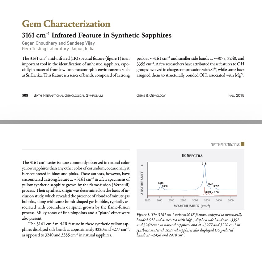
The 3161 cm⁻¹ mid-infrared (IR) spectral feature (figure 1) is an important tool in the identification of unheated sapphires, especially in material from low-iron metamorphic environments such as Sri Lanka. This feature is a series of bands, composed of a strong peak at ~3161 cm⁻¹ and smaller side bands at ~3075, 3240, and 3355 cm⁻¹. A few researchers have attributed these features to OH groups involved in charge compensation with Si⁴⁺, while some have assigned them to structurally bonded OH, associated with Mg²⁺. The 3161 cm⁻¹ series is more commonly observed in natural-colour yellow sapphires than any other colour of corundum; occasionally it is encountered in blues and pinks. These authors, however, have encountered a strong feature at ~3161 cm⁻¹ in a few specimens of yellow synthetic sapphire grown by the flame-fusion (Verneuil) process. Their synthetic origin was determined on the basis of inclusion study, which revealed the presence of clouds of minute gas bubbles, along with some bomb-shaped gas bubbles, typically associated with corundum or spinel grown by the flame-fusion process. Milky zones of fine pinpoints and a “plato” effect were also present. The 3161 cm⁻¹ mid-IR feature in these synthetic yellow sapphires displayed side bands at approximately 3220 and 3277 cm⁻¹, as opposed to 3240 and 3355 cm⁻¹ in natural sapphires.

The role of evaporites in the formation of gems during metamorphism of carbonate platforms: a review
Journal Article
Guiliani, G., Dubessy, J., Ohnenstetter, D., Banks, D., Branquet, Y., Feneyrol, J., Fallick, A.E. and Martelat, J.-E. (2018) The role of evaporites in the formation of gems during metamorphism of carbonate platforms: a review. Mineralium Deposita, Vol. 53, No. 1, pp. 1–20.
Record No. 27279  |  ISBN/ISSN: 264598  |  DOI: https://doi.org/10.1007/s00126-017-0738-4  |  Sinkankas/Born No.: NIS/NIB  |  RWHL
The mineral and fluid inclusions trapped by gemstones during the metamorphism of carbonate platform successions are precious markers for the understanding of gem genesis. The nature and chemical composition of inclusions highlight the major contribution of evaporites through dissolution or fusion, depending on the temperature of formation from greenschist to granulite facies. The fluids are highly saline NaCl-brines circulating either in an open system in the greenschist facies (Colombian and Afghan emeralds) and with huge fluid-rock metasomatic interactions, or sulphurous fluids (ruby, garnet, tsavorite, zoisite, tanzanite, and lapis-lazuli) or molten salts formed in a closed system with a low fluid mobility (ruby in marble) in the conditions of the amphibolite to granulite facies. These chloride-fluoride-sulphate ± carbonate-rich fluids scavenged the metals essential for gem formation. At high temperature, the anions SO₄²⁻, NO₃⁻, BO₃³⁻, and F⁻ are powerful fluxes which lower the temperature of chloride- and fluoride-rich ionic liquids. They provided transport over a very short distance of aluminium (Al) and/or silica (SiO₂) and transition metals which are necessary for gem growth. In summary, the genetic models proposed for these high-value and ornamental gems underline the importance of the metamorphism of evaporites formed on continental carbonate shelves and emphasise the chemical power accompanying metamorphism at moderate to high temperatures of evaporite-rich and organic matter-rich protoliths to form gem minerals.

Amethyst occurrences in tertiary volcanic rocks of greece: Mineralogical, fluid inclusion and oxygen isotope constraints on their genesis
Electronic Article
Voudouris, P., Melfos, V., Mavrogonatos, C., Tarantola, A., Gӧtze, J., Alfieris, D., Maneta, V. and Psimis, I. (2018) Amethyst occurrences in tertiary volcanic rocks of greece: Mineralogical, fluid inclusion and oxygen isotope constraints on their genesis. Minerals, Vol. 8, No. 8, 26 pp.
Record No. 17233  |  ISBN/ISSN: 2075163X  |  Sinkankas/Born No.: NIS/NIB  |  RWHL
Epithermally altered volcanic rocks in Greece host amethyst-bearing veins in association with various silicates, carbonates, oxides and sulfides. Host rocks are Oligocene to Pleistocene calc-alkaline to shoshonitic lavas and pyroclastics of intermediate to acidic composition. The veins are integral parts of high to intermediate sulfidation epithermal mineralized centers in northern Greece (e.g., Kassiteres–Sapes, Kirki, Kornofolia/Soufli, Lesvos Island) and on Milos Island. Colloform–crustiform banding with alternations of amethyst, chalcedony and/or carbonates is a common characteristic of the studied amethyst-bearing veins. Hydrothermal alteration around the quartz veins includes sericitic, K-feldspar (adularia), propylitic and zeolitic types. Precipitation of amethyst took place from near-neutral to alkaline fluids, as indicated by the presence of various amounts of gangue adularia, calcite, zeolites, chlorite and smectite. Fluid inclusion data suggest that the studied amethyst was formed by hydrothermal fluids with relatively low temperatures (~200–250°C) and low to moderate salinity (1–8 wt % NaCl equiv). A fluid cooling gradually from the external to the inner parts of the veins, possibly with subsequent boiling in an open system, is considered for the amethysts of Silver Hill in Sapes and Kassiteres. Amethysts from Kornofolia, Megala Therma, Kalogries and Chondro Vouno were formed by mixing of moderately saline hydrothermal fluids with low-salinity fluids at relatively lower temperatures indicating the presence of dilution processes and probably boiling in an open system. Stable isotope data point to mixing between magmatic and marine (and/or meteoric) waters and are consistent with the oxidizing conditions required for amethyst formation.

Archaeometric and gemmological analyses of a Roman imperial gold-and-sapphire jewel from Colonna (Rome, Italy)
Journal Article
Butini, E., Butini, F., Angle, M., Cerino, P., De Angelis, A., Tomei, N. and Altamura, F. (2018) Archaeometric and gemmological analyses of a Roman imperial gold-and-sapphire jewel from Colonna (Rome, Italy). Measurement, Vol. 128,, 2018/11/01/ pp. 160–169.
Record No. 17133  |  ISBN/ISSN: 2632241  |  DOI: https://doi.org/10.1016/j.measurement.2018.06.011  |  Sinkankas/Born No.: NIS/NIB  |  RWHL
In 2011, an extraordinary piece of gold jewelry set with precious stones was found in a woman’s tomb discovered not far from Rome and dating from the third century AD. Our examination and archaeometric analyses enabled us to identify the technique used to make the jewel and to characterize the nature and provenance of the gemstones. The analyses showed that all the stones were natural sapphires. Their inclusions and gemmological features indicate that they came from southern and south-eastern Asia, thus gaining insights about trade routes and the luxury-goods market during the Roman imperial period.

A common origin for Thai/Cambodian rubies and blue and violet sapphires from Yogo Gulch, Montana, U.S.A.?

Journal Article
Palke, A.C., Wong, J., Verdel, C. and Ávila, J. (2018) A common origin for Thai/Cambodian rubies and blue and violet sapphires from Yogo Gulch, Montana, U.S.A.? American Mineralogist, Vol. 103, No. 3, pp. 469–479.
Record No. 16850  |  DOI: https://doi.org/10.2138/am-2018-6164  |  Sinkankas/Born No.: NIS/NIB  |  RWHL
A wide number of genetic models have been proposed for volcanically transported ruby and sapphire deposits around the world. In this contribution we compare the trace element chemistry, mineral and melt inclusions, and oxygen isotope ratios in blue to reddish-violet sapphires from Yogo Gulch, Montana, U.S.A., with rubies from the Chantaburi-Trat region of Thailand and the Pailin region of Cambodia. The similarities between Thai/Cambodian rubies and Yogo sapphires suggest a common origin for gem corundum from both deposits. Specifically, we advance a model whereby sapphires and rubies formed through a peritectic melting reaction when the lamprophyre or basalts that transported the gem corundum to the surface partially melted Al-rich lower crustal rocks. Furthermore, we suggest the protolith of the rubies and sapphires was an anorthosite or, in the case of Thai/Cambodian rubies, an anorthosite subjected to higher pressures and converted into a garnet-clinopyroxenite. In this model the rubies and sapphires are rightfully considered to be xenocrysts in their host basalts or lamprophyre; however, in this scenario they are not “accidental” xenocrysts but their formation is intimately and directly linked to the magmas that transported them to the surface. The similarities in these gem corundum deposits suggests that the partial melting, non-accidental xenocryst model may be more wide-reaching and globally important than previously realized. Importantly, in both cases the gem corundum has an ostensibly “metamorphic” trace element signature, whereas the presence of silicate melt (or magma) inclusions shows they ought to be considered to be “magmatic” rubies and sapphires. This discrepancy suggests that existing trace element discriminant diagrams intended to separate “metamorphic” from “magmatic” gem corundum ought to be used with caution.

Jade Mountains and Cinnabar Pools: The History of Travel Literature in Imperial China
Book
Hargett, J.M. (2018) Jade Mountains and Cinnabar Pools: The History of Travel Literature in Imperial China. Seattle, University of Washington Press, 264 pp.
Record No. 16555  |  ISBN/ISSN: 9780300000000  |  Sinkankas/Born No.: NIS/NIB  |  RWHL
First-hand accounts of travel provide windows into places unknown to the reader, or new ways of seeing familiar places. In Jade Mountains and Cinnabar Pools, the first book-length treatment in English of Chinese travel literature (youji), James M. Hargett identifies and examines core works in the genre, from the Six Dynasties period (220–581), when its essential characteristics emerged, to its florescence in the late Ming dynasty (1368–1644). He traces the dynamic process through which the genre, most of which was written by scholars and officials, developed, and shows that key features include a journey toward an identifiable place; essay or diary format; description of places, phenomena, and conditions, accompanied by authorial observations, comments, and even personal feelings; inclusion of sensory details; and narration of movement through space and time. Travel literature’s inclusion of a variety of writing styles and purposes has made it hard to delineate. Hargett finds, however, that classic pieces of Chinese travel literature reveal much about the author, his values, and his view of the world, which in turn tells us about the author’s society, making travel literature a rich source of historical information.

The chemical component and indication of graphite inclusions in tremolite jade from Sangpiyu, Liaoning
辽宁桑皮峪透闪石玉的化学成分及石墨包裹体的指示意义
Thesis
Zhang, C. (2018) The chemical component and indication of graphite inclusions in tremolite jade from Sangpiyu, Liaoning. [辽宁桑皮峪透闪石玉的化学成分及石墨包裹体的指示意义]., Beijing, China: China University of Geosciences (Beijing), 76 pp.
Record No. 7306  |  Sinkankas/Born No.: NIS/NIB  |  Language: in Chinese with English abstract  |  RWHL
Based on the test methods such as petrography, electron probe microanalysis, cathode luminescence, X-ray powder diffraction, laser Raman spectroscopy, infrared spectroscopy, UV-visible spectroscopy and so on, the mineral composition, chemical component, spectroscopy characteristics were systematically studied in this paper and the implications for graphite inclusions were also discussed. The Sangpiyu tremolite jade shows various colours including bluish white, yellow green, deep green and dark green, although pure white variety has not yet been found. The Sangpiyu tremolite jade contains abundant graphite inclusions, mostly showing striated distribution pattern in dark green and yellow green varieties and belts of graphite in the deep green types. In dark green sample, the black belts of graphite are rather developed. Polarizing microscope reveals that the main mineral composition of the Sangpiyu tremolite jade is tremolite, with minor calcite, dolomite, zoisite, titanite, allanite, apatite and graphite. The textures of Sangpiyu tremolite jade are felt-like interweave, microfibril crystal, microscopic radial (broom) and micro-texture of crystal blade etc. The mineral grains of Sangpiyu tremolite are abundant, this suggests that tremolite jade formation process can be divided into fourth phases: platy, columnar or lamellar, fibrous and subtle fibrous. The EMPA shows that tremolite jade in Sangpiyu is predominately composed of tremolite with minor calcite, allanite, chlorite, apatite, chromite and graphite. Whereas, the major minerals of the wall rocks are plagioclase. The main chemical components of the Sangpiyu tremolite in Liaoning Province are SiO₂, MgO and CaO. Among them, the content of SiO₂ is generally more than 60%, and the content of MgO and CaO in the whole samples is slightly lower. XRD reveals that Sangpiyu tremolite is predominately composed of tremolite with minor calcite, serpentine and graphite. The graphite layers in dark green samples show a relatively constant d(002) = 0.33562, and suggest that these were formed at relatively high temperature metamorphic conditions. Based on the information of U and Rh%, it can further prove that the graphite inclusions in this kind of samples crystallize are much better. Raman spectroscopy of graphite inclusions shows that the D₁/G intensity ratio ranges from 0.78 to 0.88 in deep green samples and from 0.05 to 0.23 in dark green samples. The ranges of D₁/(D₁ + G) integral area ratio for these types are from 0.0548 to 0.3037 and 0.5528 to 0.7355 respectively. The formation temperature of graphite inclusions in the dark green tremolite jade is computed as 549.8 °C, whereas that for the deep green sample is about 343.2 °C. Our results suggest that the jade formation occurred in a multi-stage process through the action of hydrothermal fluids and metamorphism possibly in a subduction-related setting at moderate to high temperatures.

A study of gemological mineralogy of green nephrite from Qiekuotai deposit in Manasi county, Xinjiang
新疆玛纳斯切阔台碧玉的宝石矿 物学特征研究
Thesis
Liu, C. (2018) A study of gemological mineralogy of green nephrite from Qiekuotai deposit in Manasi county, Xinjiang. [新疆玛纳斯切阔台碧玉的宝石矿 物学特征研究]., Beijing, China: China University of Geosciences (Beijing), 71 pp.
Record No. 7250  |  Sinkankas/Born No.: NIS/NIB  |  Language: in Chinese with English  |  RWHL
Manasi County in Xinjiang Province has one of the most important green nephrite mines in China. The mining history can be traced back to the period of Emperor Qianlong of the Qing dynasty. The nephrite mine was found again in 1973 and later put into production. The green nephrite deposit in Manasi is the ultrabasic type of metasomatic replacement deposit. It is located in the ophiolitic mélange belt of the Early Palaeozoic in North Tianshan, Xinjiang. The nephrite is formed by the contact replacement between the ultrabasic rock and the basic country rock. It shows an occurrence of wedge-shaped or lenticular structure, where the ultrabasic rock branches intruded into the country rock. The ultrabasic rock is strongly serpentinized, and some serpentinite has been altered into talc and magnesite. The nephrite in Qiekuotai has different tones of green color, such as pale green, vivid green, or yellowish green. Some nephrite has a brown color like caramel. The green nephrite always has opaque “black spot” inclusions, or “green spot” inclusions whose color is lighter than the green nephrite. The average refractive index of nephrite is 1.61. The average relative density of nephrite is 2.96. The nephrite has a vitreous luster and ranges from opaque to translucent in transparency. It does not show an obvious absorption spectrum when observed with a spectroscope and is inert under ultraviolet light. The major mineral of the nephrite in Qiekuotai is tremolite. The textures of tremolite are felt-like blastic texture, fibrous blastic texture, harbor-like blastic texture, and coarse-grained blastic texture. The average ratio of Mg∕(Mg + Fe²⁺) is 0.934. The minor minerals are chromite, chlorite, and uvarovite. Chromite is the main component of the “black spots,” while uvarovite is the main component of the “green spots.” Analysis of the results from EMPA shows the average formation temperature and pressure of chromite are 1336.54 °C and 3.188 GPa. The average formation temperature of chlorite is 141.5 °C. The formation temperature of uvarovite is 650–350 °C. Compared with nephrite from other mines, the nephrite in Qiekuotai has less Cr content, with an average of 83.78 ppm. Also, the nephrite in Qiekuotai has a lower total amount of REE, with an average of 2.165. The nephrite samples are light REE-enriched, most with positive Eu anomalies. The nephrite samples are low in Hf, Zr, and Ta content, but enriched in Sr content. The nephrite samples have higher ratios of Rb∕Ba and Sr∕Ba. The Fe²⁺ → Fe³⁺ inter-valence charge transfer transition in tremolite causes strong visible light absorbance from 550 nm to 800 nm, and gives the nephrite its green color. The different visible light absorbance from 270 nm to 500 nm gives nephrite a tone from pale green to yellowish green, due to Fe²⁺ → O metal-to-ligand charge transfer intensity.

Preliminary study on the characteristics and quality of nephrite from Xianghualing, Hunan province

湖南香花岭软玉矿的特征与质量的初步研究
Thesis
Li, P. (2018) Preliminary study on the characteristics and quality of nephrite from Xianghualing, Hunan province. [湖南香花岭软玉矿的特征与质量的初步研究]., China: Guilin University of Technology, 42 pp.
Record No. 7241  |  Sinkankas/Born No.: NIS/NIB  |  Language: in Chinese with English abstract  |  RWHL
Hetian jade, with its rich cultural heritage, has seen changes in mining technology and geographical sources due to economic development. Apart from Xinjiang, known sources include Golmud in Qinghai, Xiuyan in Liaoning, Luodian in Guizhou, Hualien in Taiwan, and Luoyang in Henan. In 2016, a suspected nephrite deposit was discovered in TangGuanPu, XiangHuaLing area of Linwu, Hunan. The TangGuanPu area is situated at the contact zone of granite and carbonate rock in Hunan. Initially known for tin polymetallic deposits, nephrite is found as an accessory mineral. The nephrite deposits are primarily of the contact metasomatic type, resulting from contact metasomatism between granite and calcium-magnesium carbonate. Silicon-card lithified diopside, garnet, and later hydrous silicate minerals like tremolite and actinolite were formed through metasomatism. The nephrite veins in the area are about 0.5 to 1.5 meters wide, often exhibiting blue and green colors. Inclusions of quartz and metal minerals like arsenopyrite and pyrrhotite are common, affecting the transparency and appearance of the nephrite. The second mining area, being a tin polymetallic deposit, has additional minerals such as magnetic arsenopyrite and pyrite. Analyzing electron probe and X-ray fluorescence spectrum data revealed high iron content in the nephrite from the TangGuanPu area, averaging 4.366%. Other impurity elements include potassium, manganese, sodium, phosphorus, titanium, nickel, sulfur, chromium, and aluminum, contributing to uneven coloration and the presence of white veins and dark metal particles. The Mg/(Mg + Fe²⁺) ratio of 0.817 distinguishes the nephrite from Fragrant Hill as Yang nephrite, highlighting a significant difference from Xinjiang and Qinghai nephrite jade.

Exploring the Literature of Gemstone Inclusions

The literature of gemstone inclusions covers identification, origin determination, treatments, synthesis, mineral inclusions, fluid inclusions and other internal features of natural and synthetic gems.

For a curated selection of important sources, read Inclusions in Gemstones — Hyperion Literature Sources.

About Four Treasures

Four Treasures is Lotus Gemology's freely accessible gemological reference database. Use the full database to search by author, title or keyword and explore literature on gemology, mineralogy, jewelry and related fields. Where available, hyperlinks on titles may lead directly to the full publication.

Search the Complete Four Treasures Database