Gemstone Inclusions Bibliography

Gemstone Inclusions Bibliography

Explore books, articles and historical sources on inclusions in gemstones in Four Treasures, the Lotus Gemology reference database. This bibliography brings together matching records from the collection, with publication details, abstracts and cover images where available.

Gemstone Inclusion References

The list below matches the terms inclusion or inclusions in the database's searchable fields. Some references discuss inclusions as part of a broader subject.
1,001–1,020 of 1,197 matching references

Eclogites and jadeite from the Motagua Fault Zone, Guatemala
Journal Article
McBirney, A., Aoki, K.-i. and Bass, M.N. (1967) Eclogites and jadeite from the Motagua Fault Zone, Guatemala. American Mineralogist, Vol. 52, No. 5–6, May–June, pp. 908–918.
Record No. 7824  |  ISBN/ISSN: 0003004X  |  Sinkankas/Born No.: NIS/NIB  |  RWHL
Jadeite occurs as inclusions in serpentinite in the Motagua fault zone. A boulder of remarkable size and purity was found near Manzanal, Guatemala, and chemical, optical, and X-ray studies have been made of a separated sample of the jadeite. Elsewhere in the Motagua fault zone, omphacite-garnet eclogites and glaucophane-lawsonite-omphacite-garnet rocks have been found in similar environments. Only at the Manzanal locality, however, have sodic pyroxenes been found in rocks which are nearly monomineralic.

Solid inclusions in corundum and almandine garnet from Ceylon, identified by x-ray powder photographs
Journal Article
Zwaan, P.C. (1967) Solid inclusions in corundum and almandine garnet from Ceylon, identified by x-ray powder photographs. Journal of Gemmology, Vol. 10, No. 7, July, pp. 224–234.
Record No. 1924  |  ISBN/ISSN: 221252  |  Sinkankas/Born No.: NIS/NIB  |  RWHL

Determination of solid inclusions in gemstones
Journal Article
Schubnel, H.-J. (1967) Determination of solid inclusions in gemstones. Journal of Gemmology, Vol. 10, No. 6, April, pp. 189–193.
Record No. 1563  |  ISBN/ISSN: 221252  |  Sinkankas/Born No.: NIS/NIB  |  RWHL

Composition of jadeite pyroxene from the California metagraywackes
Journal Article
Coleman, R.G. (1965) Composition of jadeite pyroxene from the California metagraywackes. U.S. Geological Survey Professional Paper, No. 525-C, pp. 25–34.
Record No. 6747  |  Sinkankas/Born No.: NIS/NIB  |  RWHL
Chemical, optical and X-ray investigations demonstrate that jadeitic pyroxenes in California’s Franciscan metagraywackes contain substantial proportions of other pyroxene components and should not be treated as pure jadeite when interpreting metamorphic conditions. A purified concentrate from Angel Island, San Francisco Bay, was analysed chemically and examined for its physical properties, with comparisons drawn from Valley Ford, Pacheco Pass and previously described material from Celebes. The compared metagraywacke pyroxenes contain approximately 80–87 mol% jadeite, together with diopside, acmite and hedenbergite components. The Angel Island pyroxene has refractive indices of approximately 1.670, 1.671 and 1.678, birefringence of 0.008, strong optical dispersion and a measured density of 3.32 g/cm³, closely matching its calculated density of 3.33 g/cm³. Its unit-cell volume is slightly greater than that of nearly pure jadeite, reflecting compositional substitution. These characteristics distinguish the metagraywacke pyroxenes both from nearly pure jadeite in silica-deficient rocks enclosed by serpentine and from the less jadeitic omphacites of eclogites. The host rocks typically retain sedimentary textures and lack conspicuous schistosity, making field recognition difficult, although increased density provides a useful preliminary indication. Their characteristic assemblage comprises quartz, jadeitic pyroxene, lawsonite and muscovite, with variable glaucophane, aragonite, calcite and chlorite. Jadeitic pyroxene forms fine prismatic aggregates containing graphite and quartz inclusions. Albite is generally absent from the pyroxene-bearing groundmass but occurs in quartz veins lacking ferromagnesian minerals, suggesting that pyroxene formation involves reactions between albite and minerals such as chlorite rather than simple decomposition of albite alone. Comparisons of whole-rock analyses, sodium-to-potassium ratios and normative feldspar proportions reveal no significant chemical distinction between metamorphosed and unmetamorphosed graywackes and provide no requirement for introduction of alkalis during metamorphism. Reactions involving albite, chlorite or montmorillonite, and calcium carbonate are proposed to generate jadeitic pyroxene, glaucophane and quartz while releasing water and carbon dioxide. Incorporation of non-jadeite components may permit pyroxene formation at lower pressures than those experimentally established for pure jadeite plus quartz, but the magnitude of this effect remains uncertain and requires further investigation. Coexisting aragonite and lawsonite nevertheless support formation under high-pressure, relatively low-temperature conditions. An intermediate assemblage containing lawsonite, albite and chlorite is proposed to connect greenschist and glaucophane schist facies, with further regional mapping needed to establish their structural and metamorphic relationships.

The diagnostic significance of inclusions in precious stones
Journal Article
Eppler, W.F. (1964) The diagnostic significance of inclusions in precious stones. Lapidary Journal, Vol. 18, No. 6, September, pp. 676–679.
Record No. 719  |  Sinkankas/Born No.: NIS/NIB  |  RWHL

Sapphire from Rio Coxim, Mato Grosso, Brazil
Journal Article
Eppler, W.F. (1964) Sapphire from Rio Coxim, Mato Grosso, Brazil. Journal of Gemmology, Vol. 9, No. 6, April, pp. 199–204.
Record No. 718  |  ISBN/ISSN: 221252  |  Sinkankas/Born No.: NIS/NIB  |  RWHL
About four years ago, Professor J. E. de Souza Campos of the University of São Paulo, Brazil, reported on a new sapphire from Brazil. He mentioned strange inclusions, unknown until now in sapphire and resembling rounded gas bubbles so typical of synthetic corundum. At the author’s request, Professor de Souza Campos very kindly made available two cut stones of this sapphire and later some raw material, which could be tested more easily. The locality of these sapphires is quoted by de Souza Campos as Jauré and Quilombo, Rio Coxim, Mato Grosso. He found the refraction for sodium light to be nₒ = 1.770, nₑ = 1.762, and the birefringence 0.008. The dichroism for blue-coloured stones is o = blue to violet blue, e = green; for greenish-coloured stones o = green, e = yellow-green. With the hydrostatic balance he determined the specific gravity at 3.952 to 4.052. The stones exhibit a dark blue colour but, regrettably, the transparency is considerably reduced by many inclusions. At a first sight through the microscope, many of them indeed resembled rounded gas bubbles. But when using a higher magnification, it is seen that two kinds of inclusions are present: liquid and gas-filled discs, and negative crystals, many of them exhibiting rounded forms.

Mineralogische Untersuchungen an Granaten aus in Schweden gefundenen Schmuckgegenständen der Merowinger- und Karolingerzeit
Mineralogical investigations of garnets from jewelry items found in Sweden dating to the Merovingian and Carolingian periods
Journal Article
Mellis, O. (1963) Mineralogische Untersuchungen an Granaten aus in Schweden gefundenen Schmuckgegenständen der Merowinger- und Karolingerzeit. [Mineralogical investigations of garnets from jewelry items found in Sweden dating to the Merovingian and Carolingian periods] Arkiv för Mineralogi och Geologi, Vol. 3, No. 4, pp. 297–362.
Record No. 26791  |  Sinkankas/Born No.: NIS/NIB  |  Language: in German  |  RWHL
Mellis presents a nondestructive mineralogical investigation of 41 red stones from Merovingian- and Carolingian-period jewelry found in Sweden, undertaken both to establish whether the stones were actually garnets and, if possible, to determine their geographical origin. Because of the cultural and historical importance of the objects, the examination was restricted chiefly to refractive index, color and absorption spectra, specific gravity, anomalous birefringence, inclusions, and, for some specimens, single-crystal X-ray study. The small polished garnet plates are described individually, with detailed observations of their optical properties and inclusions, including various types of needles, prismatic and plate-like crystals, opaque minerals, zircon, and other phases. Chemical compositions were estimated from physical and optical properties and compared with established garnet compositional groups; the examined stones include pyrope-rich and almandine-rich varieties and mixtures of the principal garnet components. Mellis explores whether inclusions and composition can identify the geological environment and source of the stones, comparing the archaeological specimens particularly with garnets from Bohemia and other known occurrences. He concludes that inclusions alone cannot, at the existing state of knowledge, provide a reliable determination of geographical provenance, although inclusions combined with chemical composition can exclude certain geological environments and help characterize possible source rocks. The study demonstrates the value of detailed mineralogical examination of archaeological garnets while emphasizing the limitations of assigning ancient worked stones to specific deposits solely from their observable mineralogical properties.

Fine zoning in Australian sapphire

Journal Article
Rutland, E.H. (1963) Fine zoning in Australian sapphire. Journal of Gemmology, Vol. 9, No. 3, July, p. 83.
Record No. 1506  |  ISBN/ISSN: 221252  |  Sinkankas/Born No.: NIS/NIB  |  RWHL
Examination of a parcel of approximately thirty blue Australian sapphires reveals exceptionally closely spaced growth zoning that can resemble the fine striations associated with synthetic corundum. The stones, submitted by John Fleming of New Zealand, attracted attention because their growth bands were of a similar order of size to those seen in synthetic material. Although the observed striations showed no curvature, this distinction alone was insufficient to resolve their origin immediately: in small stones fashioned from a large synthetic boule, curvature may be inconspicuous, particularly under strong magnification. The report therefore identifies a potential difficulty in interpreting fine zoning when only a limited portion of a growth structure is visible within a faceted stone. Further microscopic examination disclosed convincingly hexagonal zoning in some specimens, together with healed fractures, providing additional evidence for their identification as natural sapphires. Photomicrographs illustrate both the closely spaced straight bands that initially prompted concern and the more diagnostic hexagonal pattern found during the subsequent investigation. Other observations document the physical and optical characteristics of the parcel. Testing in Clerici solution showed the stones to have densities slightly greater than that of a reference sapphire with a density of 3.995, although individual numerical determinations were not reported. Refractive indices measured with a Rayner refractometer in sodium light ranged from 1.765 to 1.780. Dichroism was pronounced, producing green and purple shades of the blue body colour. Examination between crossed nicols revealed neither strain nor twinning, and no further features of particular interest were recorded. The stones were described as richly blue but somewhat dark. The principal contribution is the documentation of unusually fine natural growth zoning whose scale and appearance approach those of synthetic corundum. Its diagnostic implication is that closely spaced striations should be assessed through their geometry and associated internal features, with particular attention to the possibility that a small field of observation can conceal curvature. The note offers an observational account of this identification problem rather than an explanation of the geological conditions responsible for the exceptionally narrow zones.

Corundum and amethyst from Tanganyika
Journal Article
Rutland, E.H. (1963) Corundum and amethyst from Tanganyika. Journal of Gemmology, Vol. 9, No. 4, October, pp. 132–135.
Record No. 1505  |  ISBN/ISSN: 221252  |  Sinkankas/Born No.: NIS/NIB  |  RWHL
Rutland describes corundum from newly worked deposits on the upper Umba River in Tanganyika and amethyst reportedly originating in the same country, examining specimens supplied by the gem dealer Charles Mathews following a visit in 1962. The corundum mines lie in bush country approximately fifty miles inland from Tanga, where the mineral occurs in vermiculite and can readily be separated by hand. Rough material shows little evidence of water wear and consists mainly of broken fragments, although tabular hexagonal crystals also occur. Many specimens contain internal fractures, but some display excellent transparency. Rutland suggests that the extensive area of the workings could support substantial production, without providing reserve estimates or production figures. Colours include red, blue, purple, and various intermediate shades, with frequent zoning from the outer prism faces towards the crystal centre. Examples combine blue margins with yellow interiors or pale brown margins with dark red centres. Some richly purple stones become markedly redder under tungsten illumination. Particularly strong dichroism, often visible without an instrument as the rough is rotated, produces pronounced changes such as pale pink to dark magenta or yellow to brown, creating challenges for cutters seeking the most favourable colour orientation. Microscopic observations include conspicuous twin planes, twinning visible between crossed polarizers, long irregular-edged tubes, crystal aggregates resembling those found in Burmese ruby, scattered rounded crystals, and short parallel tubes. Surface features include large and small trigons on the pinacoid and elongated ridges tentatively compared with the internal tubular structures. Measured refractive indices of approximately 1.76–1.77, estimated birefringence of 0.008, and hydrostatic specific gravities slightly below 4.00 agree with ordinary corundum properties. The amethyst was purchased in Dar es Salaam, but its precise locality could not be established; its Tanganyikan origin rests on information given to Mathews. Broken pieces and a doubly terminated crystal show richly coloured interiors enclosed by translucent milky quartz. The better material has a strong, pleasing colour with a distinct red component, carried by the ordinary ray, while the extraordinary ray appears paler and cooler. Some specimens contain abundant fine fibres identified as rutile, arranged in radiating sprays and bundles, occasionally with bent or geniculated forms. These inclusions are inconspicuous to the unaided eye except when reflected light produces a sheen. Rutland suggests that thin slices might yield attractive ornamental “hair stones.” The illustrated account records distinctive colour, growth, and inclusion features while distinguishing the documented Umba corundum locality from the uncertain provenance of the amethyst.

Testing a ruby

Journal Article
Farn, A.E. (1963) Testing a ruby. Journal of Gemmology, Vol. 9, No. 3, July, pp. 75–82.
Record No. 737  |  ISBN/ISSN: 221252  |  Sinkankas/Born No.: NIS/NIB  |  RWHL
Farn outlines a systematic approach to ruby identification, emphasizing the distinction between establishing a stone as corundum and determining whether it is natural or synthetic. Although experienced observers may recognize characteristic appearances and inclusions immediately, he argues that laboratory conclusions require corroborating tests rather than intuition or commercial reputation. Refractive-index measurements and the characteristic chromium spectrum establish ruby but do not distinguish natural from synthetic material. The spectrum is described through its red doublet, broad absorption across the yellow-green region, and fine absorption lines in the blue; Farn favours spectroscopy for small or worn stones because it avoids contact with the comparatively soft refractometer prism. Microscopic examination provides the principal evidence of origin. Features discussed include rutile silk, mineral crystals, twin planes, feathers, and the uneven colour distribution termed “treacle,” which Farn associates particularly with Burmese rubies. He contrasts these with features observed in Siamese material, including distinctive halo-like feathers surrounding opaque inclusions, while acknowledging that commercial geographical labels may follow colour and appearance rather than actual provenance. Particular attention is given to misleading similarities: natural crystals may resemble gas bubbles, synthetic bubble groups may suggest natural feathers, and internal colour effects may resemble striae in glass. Comparison specimens and repeated observation are therefore recommended. For apparently inclusion-free stones, immersion in a liquid approaching ruby’s refractive index reduces surface interference and assists the search for bubbles or curved growth structure. Farn describes rotating the specimen while adjusting focus, condenser height, diaphragm aperture, and illumination angle to reveal faint internal features. Excessive magnification may obscure slight curvature by restricting the visible length of a growth line, while focusing beneath the specimen can mistakenly bring bubbles or striae in the glass immersion cell into view. When microscopy remains inconclusive, he discusses X-ray transparency, X-ray-excited fluorescence and phosphorescence, and immersion contact photography. He regards phosphorescence following X-ray excitation as strong evidence of synthetic origin, illustrating the need for continued observation with a specimen whose afterglow appeared only after a delay. Short-wave ultraviolet fluorescence is treated more cautiously: differences in brightness may assist comparison within a parcel, but variation among natural rubies prevents its use as an independent diagnostic test. Immersion contact photography can disclose curved structure that escapes direct microscopic observation, and simple low-power examination against a diffusely illuminated white background may also prove effective. Throughout, Farn stresses the difficulty created by the closely similar optical and chemical properties of natural and synthetic ruby. His central conclusion is that careful interpretation of internal structure and inclusions, supported by complementary tests where necessary, provides the strongest basis for identification.

Ancient Egyptian Materials and Industries
Book
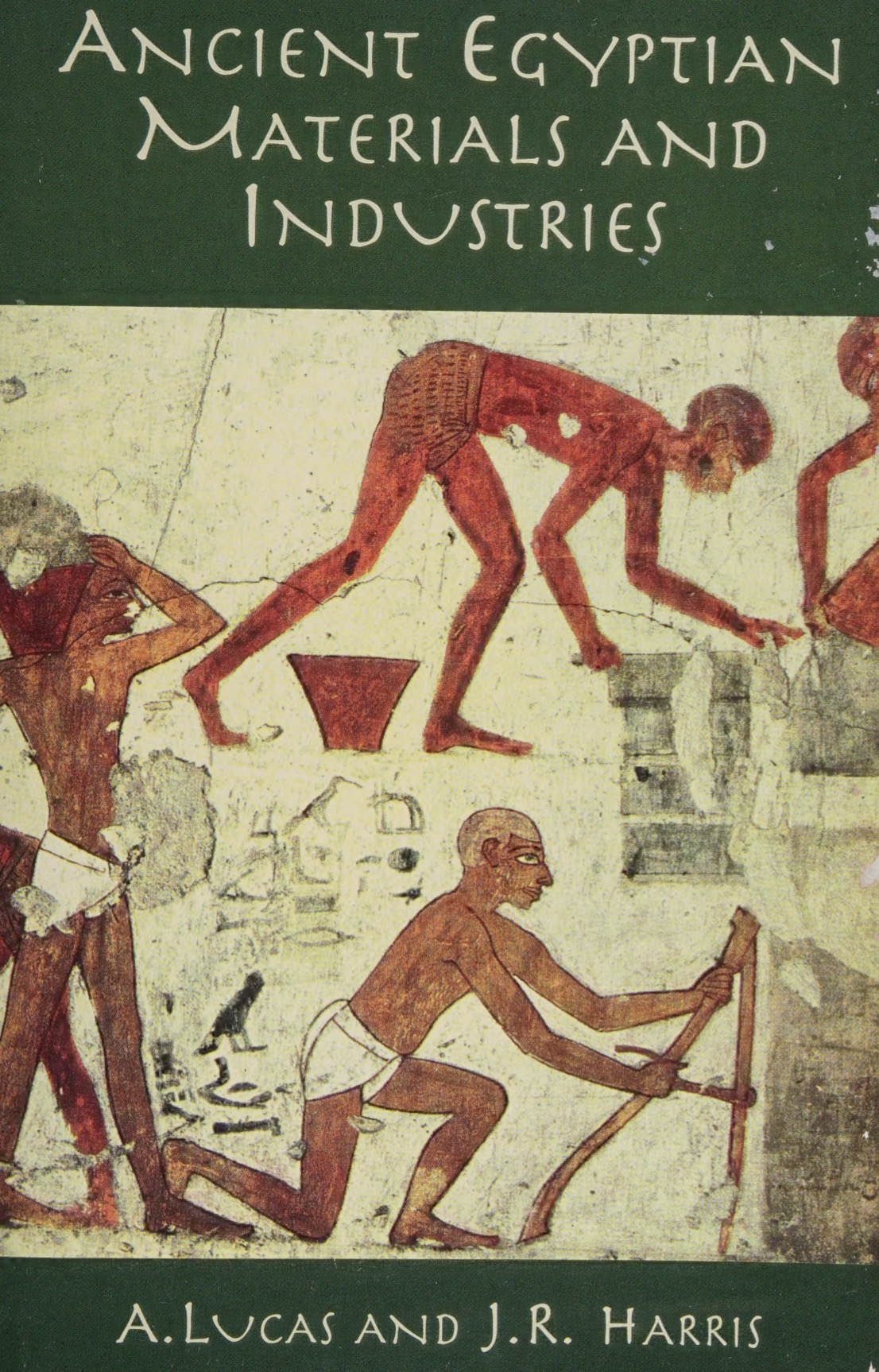
Lucas, A. and Harris, J.R. (1962) Ancient Egyptian Materials and Industries. Mineola, NY, Dover, 570 pp.
Record No. 26873  |  ISBN/ISSN: 486404463  |  Sinkankas/Born No.: 4059  |  RWHL
The majesty and splendor of ancient Egypt were largely due to the remarkably advanced technological skills developed by its artisans and craftsmen. This comprehensive and detailed study of ancient Egyptian technology meticulously documents how these workers, along with other Egyptians, exploited the land’s abundant resources. First published in 1926 and later revised and enlarged, this monumental work became a standard reference, unmatched in any other field of archaeology. The present volume reprints the fourth edition, thoroughly revised in light of more recent research and archaeological discoveries. Topics covered include a wide range of materials and techniques central to Egyptian life: the use of animal products, building materials, cosmetics, perfumes, incense, fibers, glazed ware, glass and its production, metals and alloys, painting materials, pottery, woodworking, precious and semi-precious stones, distillation of alcoholic beverages, and substances used in mummification. Profoundly erudite and rich in detail, Ancient Egyptian Materials and Industries is an indispensable reference for students of Egyptology and classical civilizations, and a vital resource for anyone interested in the long and remarkable history of technological innovation. Sinkankas: Drastically revised, rearranged: the substances now include beverages, animal products, building materials, cosmetics, perfumes & incense, ceramics & glass, fibers, metals & alloys, mummification materials, oils, fats, waxes, painting materials, pottery & its manufacture, gemstones, other stones, woods & woodworking, and a historical analysis. Ch 12, [335–54], on gemstones, among which now additionally appear coral, amber, malachite, and jade, while horn, ivory, nacre and tortoiseshell are described in Ch 2. Ornamental stones are treated in Ch 13, [355–75]. The author notes that “diamond, opal, ruby and sapphire were not known to the ancient Egyptians.” In his preface, Harris notes the inclusion of information taken from recent studies, with references added to the list in the previous edition. Much technological information is also added, with an appendix of reliable chemical analyses. “The most consistent modification has, however, been in matters of lexicography, in order to eliminate invalid evidence derived from inaccurate or disputed translations of Egyptian texts.” In this connection, see also Harris’ Lexicographical Studies in Ancient Egyptian Materials, 1961. In this edition, the following places are of gemological interest: beads, Ch 4, [41–7] (includes gemstone materials); inlaid gemstone eyes, Ch 7, [98–127] (many types & examples); gemstones in general, Ch 16, [386–405]; other stones, Ch 17, [406–28]. Lucas should always be consulted whenever one writes or lectures about Egyptian jewelry.

Testing a sapphire

Journal Article
Farn, A.E. (1962) Testing a sapphire. Journal of Gemmology, Vol. 8, No. 6, April, pp. 224–227.
Record No. 736  |  ISBN/ISSN: 221252  |  Sinkankas/Born No.: NIS/NIB  |  RWHL
Farn presents a practical account of sapphire identification, concentrating on the use of absorption spectroscopy when microscopic examination reveals no diagnostic inclusions. Drawing on routine testing experience, he describes an initial assessment of colour, cutting, and general appearance, followed by examination with a microscope or hand lens. Rotating the stone permits observation of silk, crystals, laths, twin planes, liquid feathers, two-phase inclusions, and colour zoning, which he regards as evidence of natural origin and, in favourable cases, geographical provenance. Dichroism and doubling of rear facets provide additional optical information, while refractive-index measurements establish corundum when an apparently inclusion-free specimen requires further investigation. Surface chatter or shatter marks are considered supplementary clues associated particularly with cheaply cut synthetic stones, although Farn explicitly acknowledges that they are not conclusive. The principal discussion concerns an absorption feature near 450 nanometres, which he presents as diagnostic of natural blue sapphire and absent from synthetic sapphire. His procedure begins by adjusting the spectroscope slit against daylight until the Fraunhofer lines appear sharp. A microscope with its eyepiece removed serves as an optical support, and a temporary ground-glass screen assists alignment of the lamp, specimen, and transmitted beam before the spectroscope is placed over the microscope tube. This arrangement permits examination of sapphires while still mounted in jewellery. Farn emphasizes adequate illumination and exclusion of stray light, recommending an aperture in cardboard beneath small specimens so that only light transmitted through the stone enters the instrument. He describes variation in absorption strength, contrasting the pronounced complex around 450–460 nanometres in iron-rich Australian sapphires with the weaker spectra commonly encountered in Ceylon material. Turning the specimen and slightly tilting the spectroscope may improve visibility of faint features. Practice with known natural stones is recommended to establish familiarity with the position and appearance of the spectrum before modifying the apparatus. The article also addresses heat generated by powerful lamps, noting that concentrated illumination can damage gemstones. A water-filled glass flask is proposed as both a condenser and a heat filter, while an additional flask containing copper sulphate solution suppresses the red portion of the spectrum and may enhance weak blue-region absorption. The account combines an experience-based testing sequence with detailed advice on alignment, illumination, filtering, and observation, illustrating Farn’s reliance on spectroscopy as a rapid aid to sapphire identification.

Fire marks on genuine sapphire

Journal Article
Eppler, W.F. (1962) Fire marks on genuine sapphire. Journal of Gemmology, Vol. 8, No. 5, Jan., pp. 167–170.
Record No. 716  |  ISBN/ISSN: 221252  |  Sinkankas/Born No.: NIS/NIB  |  RWHL
Eppler examines crack-like surface markings on polished corundum, commonly called fire marks or chatter marks, and challenges their widespread use as evidence of synthetic origin. Although frequently observed on synthetic ruby and sapphire, these features also occur on natural corundum and therefore cannot independently distinguish natural from laboratory-grown stones. The article reinforces Gübelin’s earlier warning that such markings reflect polishing conditions, while noting that this qualification had received insufficient attention in gemmological literature and teaching. Photomicrographs compare markings on synthetic sapphire with examples on natural sapphires of different colours and geographical origins, including sapphires from Ceylon and a greenish-blue Australian specimen. A rose-coloured Ceylon sapphire displays both surface fire marks and a healing fissure composed of small liquid inclusions. Images of chatter marks on turned steel illustrate the resemblance responsible for the alternative terminology. In metalworking, these patterns arise through displacement and overlapping of material associated with vibration, excessive loading, unsuitable turning speeds, or other operating conditions; the comparison establishes a morphological resemblance rather than an identical mechanism of formation. Eppler emphasizes the parallel arrangement of the corundum markings and their corresponding segments, together with characteristic angular relationships. Some examples show angles approaching ninety degrees, whereas others approach 120 degrees. Their appearance ranges from broad marks to fine fissure-like lines. A higher-magnification view of an Australian sapphire shows a marking continuing beneath the polished surface, supporting its interpretation as a wedge-shaped crack rather than a purely superficial blemish. Accepting excessively rapid polishing or localized overheating as the initiating cause, Eppler proposes that localized stress produces parting along planes of the primitive rhombohedron. The orientation of each polished facet relative to these crystallographic planes would then determine the directions, shapes, and angles of the resulting fissures, accounting for their varied appearance. He extends the same explanation to comparable markings on natural ruby. The article’s principal contribution is an illustrated correction to a misleading identification criterion: fire marks are polishing-related features of corundum that can occur in both natural and synthetic material, and their presence must not be treated as conclusive evidence of synthetic manufacture.

Ekanite, a new metamict mineral from Ceylon
Journal Article
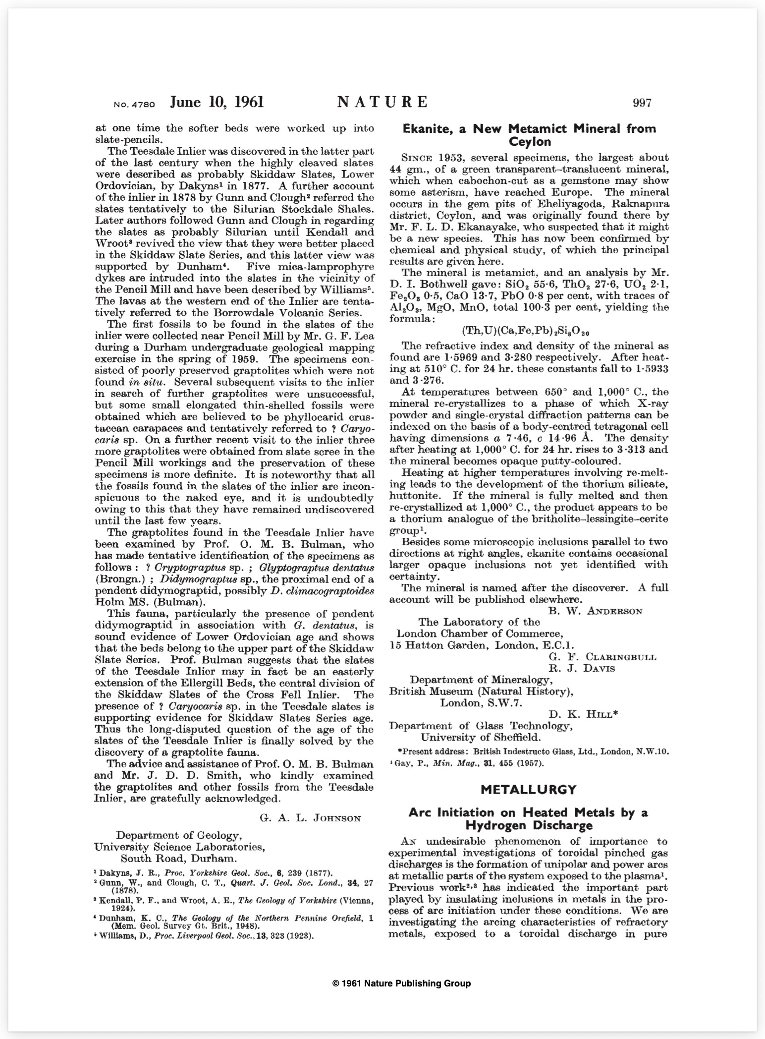
Anderson, B.W., Claringbull, G.F., Davis, R.J. and Hill, D.K. (1961) Ekanite, a new metamict mineral from Ceylon. Nature, Vol. 190, No. 4780, p. 997.
Record No. 29613  |  DOI: 10.1038/190997a0  |  Sinkankas/Born No.: NIS/NIB  |  RWHL
Anderson, Claringbull, Davis and Hill describe ekanite, a newly recognized metamict mineral from the gem pits of Eheliyagoda in the Ratnapura district of Ceylon. Known since 1953 as green transparent-to-translucent pebbles, some fashioned as cabochons showing a gemstone-quality smoky asterism, the material was recognized as unusual by F.L.D. Ekanayake, after whom the mineral was named. Chemical analysis indicates a thorium-rich calcium silicate containing uranium, lead and minor rare-earth elements, with the approximate formula (Th,U)(Ca,Fe,Pb)₂Si₈O₂₀. The mineral is metamict, with refractive index about 1.5969 and density 3.276. Heating between 650 and 1,000 °C causes recrystallization to an X-ray powder pattern resembling thorite or zircon; at higher temperatures it melts and, if subsequently recrystallized, produces an X-ray pattern analogous to huttonite. Microscopic examination shows occasional larger opaque inclusions but no identifiable inclusions responsible for the asterism.

Corundum from Tanganyika

Journal Article
Webster, R. (1961) Corundum from Tanganyika. Gems & Gemology, Vol. 10, No. 7, Fall, pp. 202–205.
Record No. 1828  |  ISBN/ISSN: 0016626X  |  Sinkankas/Born No.: NIS/NIB  |  RWHL

Hydrothermal rubies and emerald-coated beryl
Journal Article
Gübelin, E.J. (1961) Hydrothermal rubies and emerald-coated beryl. Journal of Gemmology, Vol. 8, No. 2, April, pp. 49–63.
Record No. 881  |  ISBN/ISSN: 221252  |  Sinkankas/Born No.: 2568  |  RWHL
Gübelin investigates two recently introduced gem products—rubies represented as hydrothermally grown and natural beryl coated with synthetic emerald—emphasizing the identification difficulties created when synthetic growth surrounds a natural crystal containing convincing natural inclusions. He first reviews the hydrothermal synthesis of corundum developed by Laudise and Ballman at Bell Telephone Laboratories, describing the transport of dissolved nutrient through a temperature gradient in a pressurized vessel and its deposition on suspended seed crystals. Chromium supplies the ruby colour, while growth conditions and the materials used in the apparatus influence the resulting crystals. The ruby specimens personally examined, claimed to originate from Chatham, consisted predominantly of natural ruby cores covered by relatively thin synthetic layers. Their refractive indices, birefringence, specific gravity, dichroism, absorption spectra, and fluorescence provided no reliable separation from natural ruby. The cores contained rutile silk, mineral inclusions, and liquid feathers, while the overgrowth reproduced the original crystals’ external forms. Identification depended on minute features interpreted as gas bubbles within the surface layer, occurring singly or in lines, clouds, and planes, together with the abrupt termination of natural inclusions at the boundary between core and overgrowth. Gübelin notes that ordinary cutting could remove most or all of these thin coatings. The second product, manufactured by Johann Lechleitner and marketed as Emerita, comprised a previously fashioned natural beryl nucleus overgrown with a thin hydrothermal synthetic emerald mantle. The coating continued the crystallographic orientation of the seed, forming a continuous crystal rather than a cemented assembly. Its thickness varied with crystallographic direction and controlled the apparent colour. Standard gemmological properties again overlapped those of natural emerald, and refractometer readings primarily represented the synthetic surface. Microscopy, particularly with immersion and examination from the side, revealed the dark green rim around a pale or colourless core, occasional reflections at the interface, and characteristic structures within the overgrowth. These included parallel or intersecting tension fractures, partly healed fissures containing residual liquid droplets, and particles provisionally interpreted as other mineral phases, such as phenakite or euclase. Some particles obstructed growth and produced tapering cavities containing liquid and a bubble. High magnification and phase contrast helped clarify these delicate features and their relationship to the growth process. Gübelin concludes that natural inclusions within a seed cannot establish the natural origin of the entire stone. Careful examination must instead distinguish the core from its synthetic mantle. He also stresses that altered manufacturing conditions can eliminate many diagnostic inclusions; in exceptionally clean emerald coatings, the contrast between the green outer layer and pale interior may provide the only decisive evidence.

Crystals and Crystal Growing

Book
Holden, A. and Singer, P. (1960) Crystals and Crystal Growing. Garden City, NY, Anchor Books, 320 pp.
Record No. 4646  |  Sinkankas/Born No.: NIS/NIB  |  RWHL
Holden and Singer introduce crystallography through a combination of accessible explanations, practical crystal-growing methods, and experiments that connect visible forms and physical properties with atomic organization. Written for students and amateur investigators, the book treats crystal growth as an entry into scientific inquiry, emphasizing patient observation, reproducible experiments, model building, and the relationship between evidence and explanation. Its central theme is the competition between order and disorder in matter. Discussions of solids, liquids, gases, mineral formation, ions, and salts establish a foundation for understanding repetitive atomic arrangements, crystal imperfections, and the distinction between individual crystals and polycrystalline masses. The authors then explain solutions, hydration, saturation, supersaturation, and the interpretation of solubility diagrams, showing how concentration and temperature govern crystallization. Two practical methods receive detailed treatment: growth from supersaturated solutions in sealed jars following cooling, and growth through gradual evaporation of the solvent. Instructions cover preparing solutions and seed crystals, suspending seeds, controlling growing conditions, harvesting specimens, and diagnosing difficulties such as unwanted nucleation, dissolution, rapid growth, and cloudy inclusions. Twelve recipes provide examples selected both for successful growth and for their usefulness in subsequent investigations, including alum, chrome alum, Rochelle salt, nickel sulfate hydrates, sodium chlorate, sodium bromate, sodium nitrate, and several acetate and formate compounds. The discussion then moves from growing crystals to understanding their shapes. Differential growth of faces, environmental influences, and added impurities explain variations in habit, while measurements of interfacial angles introduce crystallographic geometry. Drawings, physical models, and exercises develop the concepts of symmetry operations, crystal systems, unit cells, close packing, and atomic structures, with examples drawn from metals, salts, diamond, and related materials. Further experiments connect internal structure with cleavage, gliding, twinning, melting, and transformations between crystalline forms. Electrical behaviour is explored through piezoelectricity and its converse, while optical investigations examine double refraction, polarization, dichroism, and optical activity using comparatively simple apparatus. The final discussion extends classification beyond external shape through striations, etch figures, and the thirty-two crystal classes, then considers handed crystal structures, asymmetric molecules, and their relationship to biological substances. Appendices supply practical resources, historical context, instructions for constructing a spectroscope, symmetry tables, problems and answers, further reading, and suggestions for original investigations. Throughout, the authors make the crystals themselves experimental tools, encouraging readers to progress from producing attractive specimens to explaining how their geometry and behaviour arise from the organization of matter.

The Powder Method in X-ray Crystallography
Book
Azaroff, L.V. and Bueger, M.J. (1958) The Powder Method in X-ray Crystallography. New York, MacGraw-Hill, 342 pp.
Record No. 26929  |  Sinkankas/Born No.: NIS/NIB  |  RWHL
Instruction in the various laboratory methods for investigating crystals has long been part of the mineralogy and geology curriculum at the Massachusetts Institute of Technology, encompassing both optical and x-ray techniques. While suitable texts exist for optical methods, there has been no adequate textbook on the x-ray powder method. To support lectures and prepare students for laboratory work, the authors initially wrote a set of notes on the theory and practice of this method, which eventually evolved into this book. The expanded format allowed for more detailed discussion and the inclusion of advanced material beyond what could be covered in a short course. The powder method is inherently simple yet widely valuable across science and industry, often used by individuals with no formal training in crystallography. This book is written with such users in mind, maintaining a fundamental approach while assuming only limited prior knowledge. It omits detailed coverage of general crystallography, x-ray generation, and equipment beyond powder cameras, as these are addressed in other sources. Detection methods using photographic film are emphasized, given their simplicity and permanence, despite the growing use of ionization-type detectors. The principles outlined apply regardless of detection technique, but film is expected to remain relevant in routine applications for the foreseeable future.

Emeralds from Sandawana

Journal Article
Gübelin, E.J. (1958) Emeralds from Sandawana. Journal of Gemmology, Vol. 6, No. 8, October, pp. 340–354.
Record No. 2055  |  ISBN/ISSN: 221252  |  Sinkankas/Born No.: 2566  |  RWHL
E.J. Gübelin characterizes emeralds from the recently discovered Sandawana deposits in Southern Rhodesia [Zimbabwe], combining an account of their discovery and geological setting with chemical, optical, and microscopic examination intended to establish distinguishing features of origin. Prospectors Laurence Gontat and Cornelius Oosthuizen found the first emeralds in October 1956, followed seven months later by a richer occurrence. The deposits lie on the southern side of the Mweza Range, where pegmatite dykes intersect tremolite schists within a complex assemblage of ancient metamorphic and intrusive rocks. Gübelin interprets emerald formation as a product of contact metamorphism bringing beryllium-bearing granitic material into interaction with chromium-bearing rocks derived from ultrabasic intrusions. Study of ninety-two cut stones reveals a vivid emerald-green colour with a yellow glint, retained even in very small gems. Fracturing and small crystal size limit most available cut stones to less than a quarter carat. Chemical and spectrographic analyses indicate appreciable magnesium and sodium, together with chromium, iron, and lithium, although the small amount of material available restricts analytical accuracy. Detailed physical measurements on ten selected stones establish comparatively high refractive indices, with representative values near 1.593 and 1.586 and birefringence of approximately 0.007. Specific gravity is likewise relatively high, averaging about 2.756. The emeralds show yellow-green and bluish-green dichroism, a weak red response through the Chelsea filter, and the usual chromium-related absorption features. They are inert under both long- and short-wave ultraviolet illumination but display pale-red fluorescence in a crossed-filter fluoroscope; ultraviolet transmission ends sharply near 320 nm. Gübelin emphasizes that overlap in physical constants among emerald sources makes these measurements insufficient for reliable provenance determination by themselves. Internal characteristics provide stronger evidence, particularly short tremolite pins and exceptionally fine, sometimes curved fibres occurring singly or in bundles. Their morphology and distribution are compared with amphibole inclusions in Habachtal emeralds. Additional features include partly resorbed garnets with brownish halos, limonite-filled cracks, rare hematite, feldspar and magnetite inclusions, dustlike patches, and distinctive systems of fine fissures parallel to the basal plane. Lace-like residual liquid films observed in one stone are recorded without being treated as an established locality characteristic. Photomicrographs and comparative tables support the conclusion that Sandawana emeralds combine attractive colour with a recognizable suite of internal features useful in distinguishing them from other contemporary sources.

Notes on asterism in corundum, rose quartz and almandine garnet and chatoyancy in beryl

Journal Article
Eppler, W.F. (1958) Notes on asterism in corundum, rose quartz and almandine garnet and chatoyancy in beryl. Journal of Gemmology, Vol. 6, No. 5, January, pp. 195–212.
Record No. 715  |  ISBN/ISSN: 221252  |  Sinkankas/Born No.: NIS/NIB  |  RWHL
W.F. Eppler examines the inclusions responsible for asterism in spinel and chatoyancy in chrysoberyl, quartz, tourmaline, zircon, and scapolite, emphasizing the need to distinguish solid crystals from apparently similar hollow channels and to assess whether inclusions were trapped during growth or formed subsequently by exsolution. In natural spinel, oriented rutile needles following octahedral edges provide one explanation for asterism, but other fibrous inclusions remain unidentified. Sphene crystals in Ceylon spinel illustrate the distinction between relatively large primary crystals enclosed during host growth and minute needles attributed to exsolution. Partial dissolution and the arrangement of larger crystals on former growth planes support their primary origin, whereas their sparse distribution prevents them from producing a star. Burmese spinels contain additional unidentified dark crystals and minute brownish particles tentatively interpreted as rutile; patterned concentrations around larger inclusions suggest localized strain may influence exsolution. Experiments with synthetic spinel show that heat-induced separation of excess alumina can generate oriented plates and weak asterism, but the resulting effects lack commercial value. Elongated gas inclusions can also produce a four-rayed star in suitably oriented cabochons. Examination of a sharply chatoyant chrysoberyl reveals short, birefringent needles with a refractive index higher than the host, contradicting an explanation based exclusively on hollow channels for that specimen. Their identity and origin remain uncertain, although exsolution is suggested. Similarly, inclusions in a rose-coloured tourmaline that resemble channels in reflected light prove in transmitted light to be brown, birefringent crystal fibres with a higher refractive index than tourmaline. Eppler interprets these as primary inclusions formed alongside the host. Quartz cat’s-eye and tiger’s-eye are discussed in terms of aligned amphibole-asbestos fibres, while an earlier observation of zircon chatoyancy is attributed to unidentified minute crystals parallel to its principal axis. Madagascar scapolite displays a combination of hollow and liquid-filled channels, crystalline needles, and oriented blades, all potentially contributing to its cat’s-eye effect. The blades are interpreted as products of exsolution within the marialite–meionite series. Photomicrographs demonstrate that similar optical phenomena can arise from inclusions differing in composition, physical state, morphology, and developmental history, making detailed optical examination essential to explaining individual specimens.

Exploring the Literature of Gemstone Inclusions

The literature of gemstone inclusions covers identification, origin determination, treatments, synthesis, mineral inclusions, fluid inclusions and other internal features of natural and synthetic gems.

For a curated selection of important sources, read Inclusions in Gemstones — Hyperion Literature Sources.

About Four Treasures

Four Treasures is Lotus Gemology's freely accessible gemological reference database. Use the full database to search by author, title or keyword and explore literature on gemology, mineralogy, jewelry and related fields. Where available, hyperlinks on titles may lead directly to the full publication.

Search the Complete Four Treasures Database