Gemstone Inclusions Bibliography

Gemstone Inclusions Bibliography

Explore books, articles and historical sources on inclusions in gemstones in Four Treasures, the Lotus Gemology reference database. This bibliography brings together matching records from the collection, with publication details, abstracts and cover images where available.

Gemstone Inclusion References

The list below matches the terms inclusion or inclusions in the database's searchable fields. Some references discuss inclusions as part of a broader subject.
461–480 of 1,247 matching references

Micro-World: Fissure with moiré pattern in spinel
Journal Article
Smith, T. (2023) Micro-World: Fissure with moiré pattern in spinel. Gems & Gemology, Vol. 59, No. 3, Fall, pp. 375–376.
Record No. 6780  |  ISBN/ISSN: 0016626X  |  Sinkankas/Born No.: NIS/NIB  |  RWHL
Surface-reaching fissures commonly host precipitates of epigenetic minerals, as was the case with a 4.22 ct purple spinel recently examined by the author. “Islands” of unidentified birefringent inclusions occupied a near-planar fissure. These inclusions were inert to Raman spectroscopy. Delicate depositions radiating from these islands interacted to create a complex moiré pattern (see above). Moiré patterns, named after their resemblance to a type of fabric, form when parallel or concentric lines overlap. It is unclear whether the islands formed first and were partially dissolved in a secondary event, or if they formed simultaneously with the spinel. Although moiré patterns have been observed in partially healed fluid fingerprints and surface-reaching fissures, it is rare to see them expressed in such a spectacular form.

Lab Notes: “Nebula” inclusion in ruby beryllium-diffused to heal fractures
Journal Article
Damba, M. (2023) Lab Notes: “Nebula” inclusion in ruby beryllium-diffused to heal fractures. Gems & Gemology, Vol. 59, No. 3, Fall, pp. 367–368.
Record No. 6778  |  ISBN/ISSN: 0016626X  |  Sinkankas/Born No.: NIS/NIB  |  RWHL
Recently the Carlsbad laboratory received a 4.12 ct purple-red stone measuring approximately 11.50 × 7.70 × 4.72 mm (figure 1) for identification services. It featured unusual veil-like reddish color zoning wafting throughout a purple bodycolor. The refractive index measured 1.760–1.770, identifying the stone as a ruby. Microscopic examination indicated that the reddish color zoning was associated with altered fingerprints that resembled a cosmic nebula due to a flux-rich residue (figure 2). These fingerprints appeared to be consistent with fractures that had been healed using a flux-assisted heating process in a high-heat environment. Due to the indications of high heat, laser ablation–inductively coupled plasma–mass spectrometry (LA-ICP-MS) chemical analysis was performed to check for the presence of beryllium diffusion. Initially, LA-ICP-MS conducted on two spots revealed beryllium concentrations of <0.019 ppma and 60.87 ppma. This significant discrepancy prompted additional LA-ICP-MS testing in three spots, revealing beryllium concentrations of <0.19 ppma, 1780 ppma, and <0.19 ppma. Microscopic observation of the testing spots showed areas of high beryllium on or near the reddish color zones, while areas of very low beryllium were associated with testing areas on the purple areas of the stone. Therefore, it was concluded that a beryllium-rich flux was used to heal the fractures naturally present in the stone. The ruby was likely heated in this flux at temperatures high enough to allow the beryllium to seep from the flux into the stone, but not long enough for the beryllium to infiltrate its entirety.

Analysis of the causes of colours in blue and green chalcedony
Journal Article
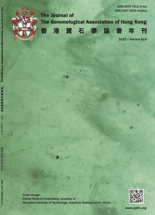
Guo, J., Liao, R. and Wang, L. (2023) Analysis of the causes of colours in blue and green chalcedony. Journal of the Gemmological Association of Hong Kong, Vol. 44, pp. 29–38.
Record No. 6647  |  Sinkankas/Born No.: NIS/NIB  |  Language: in Chinese with English abstract  |  RWHL
This paper presents research carried out on 10 samples of blue and green chalcedony to analyse and further discuss the cause of their colour. The gemmological characteristics of the samples are analysed using standard gemmological testing, energy-dispersive spectroscopy (EDS) testing, infrared spectroscopy testing, Raman spectroscopy and ultraviolet–visible spectroscopy testing. The results are as follows: The colour of the samples is mainly blue and green, with obvious shades accompanied by auxiliary tones such as yellow and grey; the spot measurement refractive index is 1.53–1.54; density is 2.59–2.63 g/cm³; fluorescence is inert; under crossed polarisers, the samples all show bright and extinction; under magnification, the structure is observed to be fine, with floccule-like inclusions, dark inclusions and watermark-like inclusions (Ed. Similar to left 3rd set of photos in p. 83 of 2022 GAHK Journal). The infrared spectrum shows the vibration peaks of typical quartzite; in addition to the quartz vibration peak, the Raman spectrum also shows the characteristic peak of 500 ± cm⁻¹ clinosilicate, and the dark blue-green sample also has a weak characteristic peak of chrysocolla. X-ray energy spectrometer (EDS) and ultraviolet–visible spectrum analysis further confirmed that the cause of colour in the light green sample was Ni; in the light blue, light grey-green, light yellow-green and dark blue-green samples it was Cu, with the dark bluish-green colour also caused by silicon impurities of chrysocolla; the yellowish-green and dark green samples were all artificially dyed by Cr salt.

G&G Microworld: Pink sapphire with graphite inclusions
Journal Article
Hernandez, J. (2023) G&G Microworld: Pink sapphire with graphite inclusions. Gems & Gemology, Vol. 59, No. 2, Summer, p. 227.
Record No. 6551  |  ISBN/ISSN: 0016626X  |  Sinkankas/Born No.: NIS/NIB  |  RWHL
Describes a graphite crystal in a pink sapphire said to be from Sri Lanka.

Gemological and chemical composition characteristics of basalt-related rubies from Chanthaburi-Trat, Thailand
Journal Article
Li, E. and Xu, B. (2023) Gemological and chemical composition characteristics of basalt-related rubies from Chanthaburi-Trat, Thailand. Crystals, Vol. 13, No. 8, p. 1179, 17 pp..
Record No. 6541  |  ISBN/ISSN: 20734352  |  Sinkankas/Born No.: NIS/NIB  |  RWHL
The geographic origin determination of ruby is increasingly important in the gem trade and geological research. Unlike metamorphic-related ruby, the rarer basalt-related ruby has gained significant attention, especially from Thailand, a major producer of such gems. Thai rubies are classified as magmatic-related origin rubies, which can be found as xenocrysts (xenoliths) hosted by alkali basalts. This paper focuses on the gemological characteristics, inclusion morphology, identification, and geochemistry of basalt-hosted ruby from the Chanthaburi–Trat area in Thailand. Various instruments, including conventional gemological tools, Raman spectrometer, EPMA, and LA-ICP-MS, were used for analysis. This study aimed to identify the distinctive characteristics of rubies from Thailand and to find feasible methods for their geographic origin determination in comparison with rubies from Cambodia, Myanmar, and Mozambique. Thailand samples exhibit diverse inclusion scenes and contain a variety of crystal or mineral inclusions. Raman spectroscopy results indicate the presence of anorthite, titanium oxide, and gypsum inclusions. The main chemical composition of the ruby consists of Al₂O₃, with trace elements including Fe, Cr, Si, Mg, Ti, Ga, V, Ca, and Ni. The color of Thailand ruby is correlated with the content of Cr and Fe. Chemical diagrams illustrating the contents of Fe, Mg, Cr, V, Ti, and Ga offer reasonable discrimination tools for differentiating rubies from various deposit types. The chemical compositions and inclusion characteristics of rubies from Thailand serve as reliable indicators for their origin identification. This study is an advantageous supplement to the research on Thailand rubies.

Gem News International: Heated purplish pink sapphire from Ilakaka (Madagascar) with colored monazite inclusions
Journal Article
Hennebois, U., Delaunay, A. and Karampelas, S. (2023) Gem News International: Heated purplish pink sapphire from Ilakaka (Madagascar) with colored monazite inclusions. Gems & Gemology, Vol. 59, No. 1, Spring, pp. 159–160.
Record No. 6537  |  ISBN/ISSN: 0016626X  |  Sinkankas/Born No.: NIS/NIB  |  RWHL

Gem News International: Forsterite in a purple Tanzanian spinel
Journal Article
Wu, J., Ma, H. and Li, H. (2023) Gem News International: Forsterite in a purple Tanzanian spinel. Gems & Gemology, Vol. 59, No. 1, Spring, pp. 150–152.
Record No. 6536  |  ISBN/ISSN: 0016626X  |  Sinkankas/Born No.: NIS/NIB  |  RWHL
Brief gemological description of low-grade ruby from Liberia.

Micro-World: Unusual solid inclusions in flame-fusion ruby
Journal Article
Kiyak, E. (2023) Micro-World: Unusual solid inclusions in flame-fusion ruby. Gems & Gemology, Vol. 59, No. 1, Spring, pp. 89–90.
Record No. 6531  |  ISBN/ISSN: 0016626X  |  Sinkankas/Born No.: NIS/NIB  |  RWHL
The author recently examined unusual dark red solid inclusions in a flame-fusion laboratory-grown ruby. Standard gemological testing yielded a refractive index of 1.760–1.768, a chromium emission line in red using a handheld spectroscope, medium red fluorescence under long-wave UV, and weak red fluorescence under short-wave UV. These properties were consistent with ruby. Microscopic examination using brightfield illumination revealed numerous dark red inclusions (figure 1) that resembled crystals commonly seen within natural corundum. Furthermore, magnification showed characteristic internal features of flame-fusion ruby such as curved striae and gas bubbles. The strong curved growth lines and numerous gas bubbles (figure 2) suggested that this was an early type of flame-fusion ruby. Although flame-fusion corundum is widely available in the marketplace, it is unusual to encounter a specimen containing natural-looking dark red solid inclusions. These inclusions are possibly unmelted alumina powder, and the fact that Raman was unable to identify something other than alumina supports this (E.J. Gübelin and J.I. Koivula, Photoatlas of Inclusions in Gemstones, Volume 3, Opinio Publishers, Basel, Switzerland, 2008). Identification of the natural or laboratory-grown origin of a stone with this type of natural-looking inclusion can be difficult. Careful observation is needed to correctly identify such a specimen’s origin.

Micro-World: Arrow-patterned negative crystals in sapphire
Journal Article
Siritheerakul, P. and Hei, T.S. (2023) Micro-World: Arrow-patterned negative crystals in sapphire. Gems & Gemology, Vol. 59, No. 1, Spring, p. 89.
Record No. 6530  |  ISBN/ISSN: 0016626X  |  Sinkankas/Born No.: NIS/NIB  |  RWHL
Negative crystals are a common inclusion in corundum and often rupture upon exposure to moderate heat treatment. The author recently analyzed a sapphire containing a series of negative crystals arranged in an arrow-like pattern (see above). Careful examination revealed a partially healed fissure intersecting one of the negative crystals, indicating the stone had been subjected to heat treatment. Fortunately, this spectacular pattern of negative crystals endured the heat treatment process, decorating the internal micro-world of this sapphire.

Lab Notes: Ruby with interesting synthetic overgrowth
Journal Article
Ahline, N. (2023) Lab Notes: Ruby with interesting synthetic overgrowth. Gems & Gemology, Vol. 59, No. 1, Spring, pp. 81.
Record No. 6528  |  ISBN/ISSN: 0016626X  |  Sinkankas/Born No.: NIS/NIB  |  RWHL
The Carlsbad laboratory received a 3.02 ct ruby for a colored stone identification and origin report. Standard gemological testing was consistent with ruby: a hydrostatic specific gravity of 3.96 and a ruby spectrum in a handheld spectroscope. Internally, the stone displayed white flakes in a hexagonal formation and roiled graining. Partially healed fissures with residue, which result from heating in the presence of a flux, were also documented. The inclusion scene, along with the stone’s trace element chemistry collected using laser ablation–inductively coupled plasma–mass spectrometry (LA-ICP-MS), were consistent with Burmese ruby. This ruby was not treated by beryllium diffusion, as no beryllium was detected by LA-ICP-MS. The curious feature of this stone was a large cavity on the pavilion that was partially filled with synthetic overgrowth that was observed in the microscope (see above). Synthetic overgrowth can be a byproduct of heating a piece of corundum at high temperatures, with or without the presence of a flux. This creates an environment in the crucible that allows for the partial dissolution of corundum and can result in synthetic corundum overgrowth on the surface and in cavities of the heated stone. Synthetic overgrowth is typically an aggregated structure of hexagonally shaped, platy crystals (Fall 2002 G&G Lab Notes, pp. 255–256). When viewed in reflective lighting, there is no luster difference between the host corundum and the synthetic overgrowth, as the two materials have the same refractive index. The gaps that could be observed between the crystals in this stone showed how they grew in different orientations. Synthetic overgrowth is common enough that it should always be looked for in corundum that has undergone high-temperature heat treatment. There are cases where synthetic overgrowth can be observed using polarized light, but careful analysis is required because the overgrowth can completely fill cavities and be inconspicuous.

Libyan desert glass bangle bracelet

Journal Article
Sin, C.Y. (2023) Libyan desert glass bangle bracelet. Gems & Gemology, Vol. 59, No. 1, Spring, pp. 73–74.
Record No. 6527  |  ISBN/ISSN: 0016626X  |  Sinkankas/Born No.: NIS/NIB  |  RWHL
Libyan Desert glass is a light green or yellow natural glass which is composed of almost 98% silica. It is a type of tektite formed by meteorite impact in the desert and the fast quenching of the silica-rich melt produced. It was first reported by Clayton and Spencer in 1934 (F. Fröhlich et al., “Libyan Desert Glass: New field and Fourier transform infrared data,” Meteoritics and Planetary Science, Vol. 48, No. 12, 2013, pp. 2517–2530) and is found in Egypt’s Western Desert. Common inclusions are bubbles, cristobalite, dark brown streaks, and black iron oxides (J.A. Barrat et al., “Geochemistry and origin of Libyan Desert glasses,” Geochimica et Cosmochimica Acta, Vol. 61, No. 9, 1997, pp. 1953–1959). Recently, GIA’s Hong Kong laboratory examined a transparent light yellow bangle bracelet measuring 66.12 × 13.10 mm and weighing 144.67 ct (figure 1). It had a spot refractive index of 1.44 and revealed weak yellow fluorescence in short-wave UV. The chemical composition, determined by qualitative analysis using energy-dispersive X-ray fluorescence, was nearly pure silica with minor amounts of iron, strontium, potassium, titanium, and zirconium. Based on the differences in molecular vibrations detected with Fourier-transform infrared (FTIR) analysis, it was determined that the molecular structure of Libyan Desert glass is significantly different from that of other pure silica glasses. It has a higher ratio of discontinuities and defects in the tetrahedral (SiO4) network (F. Fröhlich et al., 2013). Microscopic observation revealed common inclusions in glass such as numerous gas bubbles (figure 2) and flow structure. This bangle also presented abundant white spherulites identified by Raman spectroscopy as cristobalite (a polymorph of silica). These spherulites were small and individually dispersed throughout the glass (figure 3). The presence of this high-temperature low-pressure silica phase indicates a natural high-temperature formation. From all the gemological, FTIR, and Raman data collected, this bangle was identified as Libyan Desert glass. Most Libyan Desert glass is tubular in shape, and some are approximately spherical or rod-like (R.A. Weeks et al., “Libyan Desert glass: A review,” Journal of Non-Crystalline Solids, Vol. 67, No. 1-3, 1984, pp. 593–619). Those fragments are usually 2 to 63 mm in size, so it is surprising to examine this material in the form of a bangle bracelet.

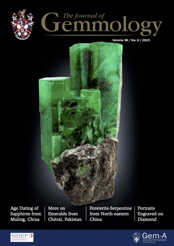
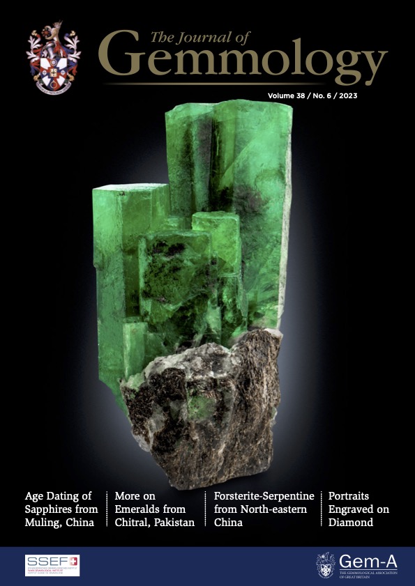
Characterisation and in situ age determination of zircon inclusions in sapphires from Muling, north-east China
Journal Article
Liu, Y., Lu, R. and Pan, S. (2023) Characterisation and in situ age determination of zircon inclusions in sapphires from Muling, north-east China. Journal of Gemmology, Vol. 38, No. 6, pp. 564–581.
Record No. 6516  |  ISBN/ISSN: 221252  |  DOI: https://doi.org/10.15506/JoG.2023.38.6.564  |  Sinkankas/Born No.: NIS/NIB  |  RWHL
In recent decades, blue and fancy-colour sapphires have been gathered from secondary deposits derived from Tertiary basalts in the Muling area of north-eastern China. In this article, zircon inclusions in the blue sapphires were studied to characterise their morphology and structure, as well as to obtain the age of the host sapphires. The zircon inclusions in the studied samples were euhedral and transparent, and their dominant morphology was similar to that of typical zircon inclusions in sapphires associated with alkali basalts. Raman spectroscopy of the zircons showed only slight radiation damage, and cathodoluminescence images revealed two distinctive zoning patterns (in their cores and rims). LA-ICP-MS analyses of the zircon rims showed typical magmatic Th/U ratios. In situ U-Pb dating of the zircon rims yielded ages of 9 to 8 Ma—which is the inferred upper limit of the formation age of the host sapphires—while cores had much older ages that exceeded 110 Ma (i.e. an inherited age component). The 9–8 Ma sapphire age coincides with Late Miocene basaltic eruptions in the Dunhua-Mishan graben. Age dating of zircon inclusions in the Muling sapphires helps elucidate their geological origin and provides useful information for the provenance determination of gem corundum.

Gem Notes: Polymer carving imitating dickite
Journal Article
Tay, T.S., Franz, L., Ling, K.H., Heng, E.C. and Ying, L.H. (2023) Gem Notes: Polymer carving imitating dickite. Journal of Gemmology, Vol. 38, No. 6, pp. 559–561.
Record No. 6514  |  ISBN/ISSN: 221252  |  DOI: https://doi.org/10.15506/JoG.2023.38.6.600  |  Sinkankas/Born No.: NIS/NIB  |  RWHL
The authors recently encountered a yellow carving (Figure 27) resembling dickite or perhaps talc, such as documented for another piece from the same collec- tion (Tay et al. 2023). However, this one turned out to be much different. When handled, the 3,550.6 g carving seemed rather heavy for its size and had a plastic-like feel. When examined with a 10× loupe, small rounded beads were visible along grooves in the carving (Figure 28), and some parts showed specks of white inclusions. The spot RI reading was 1.55, and unlike dickite, the carving fluoresced weak chalky blue to a long-wave UV lamp (although it was inert to short-wave UV radiation).

Unique interstitial textures within coarse-grained jadeitite from Kazakhstan and their significance in locality identification
Journal Article
Wen, J., Shi, G., Xing, B., Long, T. and Zhang, J. (2023) Unique interstitial textures within coarse-grained jadeitite from Kazakhstan and their significance in locality identification. Minerals, Vol. 13, No. 4, 16 pp..
Record No. 6462  |  ISBN/ISSN: 2075163X  |  Sinkankas/Born No.: NIS/NIB  |  RWHL
Unique finer-grained interstitial textures, occurring as small blocks or irregular shapes measuring 0.15–10 mm, were discovered merging into the coarse-grained textures of Kazakhstan jadeitite. Based on mineral content, these interstitial textures are categorized into two types: Type I, composed almost entirely of jadeite crystals with minor omphacite and trace analcime; and Type II, dominated by omphacite and analcime with only minor jadeite. Both types exhibit no significant preferred orientation, have sharp boundaries with the surrounding coarse textures, and display increased transparency, finer grain size, and higher idiomorphism. The coarse-grained textures include granitoid structures and radial clusters. Granitoid textures are formed by euhedral to subhedral prismatic crystals showing rhythmic zoning and parallel intergrowths. Fluid inclusions containing H₂O and CH₄ suggest that these textures resulted from the precipitation of jadeitic fluids. However, due to either limited fluid supply or excess available space, some voids remained between the coarse jadeitite grains. These voids were later filled by precipitation from later H₂O-rich fluids under different pressure–temperature conditions, producing the two types of interstitial textures. Such textures appear unique to Kazakhstan and may serve as a distinguishing visual feature for identifying Kazakhstan jadeitite.

A comparative study of the gemological characteristics and inclusions in spinels from Myanmar and Tajikistan
Electronic Article
Zhang, Y., Zhu, J.-R. and Yu, X.-Y. (2022) A comparative study of the gemological characteristics and inclusions in spinels from Myanmar and Tajikistan. Crystals, Vol. 12, No. 5, 14 pp.
Record No. 29219  |  ISBN/ISSN: 2073-4352  |  Sinkankas/Born No.: NIS/NIB  |  RWHL
Currently, most of the popular spinels in the jewellery market come from Myanmar and Tajikistan. It is well known that provenance is one of the main factors affecting the value of a gemstone, and the geographic origin of a gemstone can be determined by examining its gemological and inclusion characteristics. This study systematically characterized the conventional gemology of spinels from Myanmar and Tajikistan and compared the inclusions in the spinels from these two countries by means of gemological microscopy and Raman spectroscopy. The results showed that most red and pink Myanmarese spinels were octahedral or contact twins, while Tajikistan spinels are slabbed or octahedral distorted crystals. Columnar zircon is frequently found in Tajikistani spinels but rare in Myanmar spinels, appearing as tiny accessory inclusions. There are three types of carbonate inclusions (magnesite, dolomite, and calcite) in Myanmar spinels, but Tajikistan spinels have only one (magnesite). In addition, spinels of different origins include special inclusions. Myanmar spinels contain pyrite inclusions; Tajikistan spinels contain rutile and talc inclusions.

Origin of blue-water jadeite jades from Myanmar and Guatemala: Differentiation by non-destructive spectroscopic techniques
Journal Article
Zhang, Y. and Shi, G. (2022) Origin of blue-water jadeite jades from Myanmar and Guatemala: Differentiation by non-destructive spectroscopic techniques. Crystals, Vol. 12, No. 10,
Record No. 6658  |  ISBN/ISSN: 20734352  |  Sinkankas/Born No.: NIS/NIB  |  RWHL
Identifying the origin of jadeite jades has become increasingly important from both mineral resource and metamorphic geology perspectives. In this study, we differentiate Myanmar gem-quality blue-water jadeite jades from their Guatemala counterparts via integrating various non-destructive spectrographic techniques, including X-ray fluorescence (XRF), Ultraviolet–Visible spectroscopy (UV–Vis), Fourier transform infrared spectroscopy (FTIR), and Raman spectroscopy. Our results show that the Myanmar blue-water jadeite jades are structurally homogenous with very few impurities, while their Guatemala counterparts commonly have a yellowish margin with scattered white albite and disseminated greenish inclusions of omphacite and (minor) aegirine–augite. Geochemically, the UV absorption spectral data indicate that the Guatemala samples have higher total Fe and Fe²⁺ contents, but lower Fe³⁺ content than the Myanmar samples. The Guatemala samples also have higher omphacite content (lower molar Na/(Na+Ca) ratio, as reflected by the lower IR absorption peak wavenumber) and higher heterogeneity (as reflected by the ~680 cm⁻¹ Raman peak shift difference) than that from Myanmar. Major differences are also discovered in the blue series (Myanmar: 0–0.7 cm⁻¹; Guatemala: 1.7–3.2 cm⁻¹) and blue-green series (Myanmar: 6.9 cm⁻¹; Guatemala: 13.7 cm⁻¹) of the Raman peak shift difference, which altogether can provide a novel, nondestructive method for distinguishing blue-water jadeite jades from different origins.

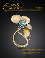
A new model for the formation of nephrite deposits: A case study of the Chuncheon nephrite deposit, South Korea
Journal Article
Feng, Y., He, X. and Jing, Y. (2022) A new model for the formation of nephrite deposits: A case study of the Chuncheon nephrite deposit, South Korea. Ore Geology Reviews, Vol. 141,, 2022/02/01/ 14 pp..
Record No. 6476  |  ISBN/ISSN: 1691368  |  DOI: https://doi.org/10.1016/j.oregeorev.2021.104655  |  Sinkankas/Born No.: NIS/NIB  |  RWHL
The Chuncheon nephrite deposit is the largest in South Korea with an estimated reserve of 300,000 tons. Located in mid-eastern South Korea, the deposit is in the form of lenses in the contact zone between dolomitic marble and amphibole schist in the upper part of a biotite schist. To date, the origin, process of formation, and age of the Chuncheon nephrite deposit remain unclear, which hinders our further understanding of the formation of nephrite. This study involved petrography, chemical analyses, and nephrite age determinations using optical mineralogy, electron microprobe analyses (EMPA), X-ray fluorescence spectroscopy (XRF), and SHRIMP (Sensitive High-Resolution Ion MicroProbe) analyses of zircon. Petrographic studies indicated that the Chuncheon nephrite consists mainly of tremolite. The low contents of Cr (2.15–4.74 ppm) and Ni (2.95–4.90 ppm) are typical of dolomite-related nephrites and lower than in serpentine-related nephrites. The total REE contents are <18.5 ppm. The REE chondrite-normalized patterns display right-leaning LREE and almost flat HREE patterns, which are consistent with the REE model of dolomitic marble. The δ¹⁸O isotope compositions of the zircons (6.34‰–12.5‰) from the nephrite overlap those of the Chuncheon dolomite (2.4‰–18.2‰), indicating clearly that dolomite played an important role in the formation of the Chuncheon nephrite. The petrography and geochemistry suggest that it had a metasomatic origin involving the reactions dolomitic marble → diopside, diopside → tremolite, and tremolite → chlorite. The δD and δ¹⁸O isotope compositions of our specimens of nephrite (δD = −95.3‰ to −78.3‰ and −118‰ to −105‰; δ¹⁸O = −9.9‰ to −7.9‰) and the hydrothermal fluids in equilibrium with the nephrite (δD = −73.6‰ to −56.6‰ and −96.3‰ to −83.3‰, 330–450 °C; δ¹⁸O = −9.41‰ to −6.38‰, 330–430 °C) suggest that the hydrothermal fluids were predominantly meteoric water. The zircons contain almost no mineral inclusions, which is quite different from zircons in other nephrite deposits. The variable SHRIMP U–Pb ages of the zircons (908.9 ± 4.8 to 459.1 ± 1.9 Ma (n = 7), 436.3 ± 16.9 to 359.5 ± 6.4 Ma (n = 5), and 249.9 ± 5.2 to 79.4 ± 1.9 Ma (n = 5)) in the Chuncheon nephrite may record the ages of a regional multi-stage metamorphism. Due to the generally low temperature of formation of the nephrite, we speculate that the ages of zircons only record the two relatively high-temperature initial stages of nephrite formation (dolomitic marble → diopside, and diopside → coarse-grained tremolite) and failed to record the age of the lowest-temperature third stage of high-quality nephrite formation (coarse-grained tremolite → fine-grained tremolite). It has generally been considered that dolomite-related nephrite forms at the contacts between granite/granodiorite and dolomitic marble, whereas serpentinite-related nephrite forms at the contacts between serpentinite and silica-rich rocks. However, the genesis of the Chuncheon nephrite deposit does not fit with either of these well-known types. Thus, we developed a new model for the Chuncheon nephrite in which heat was provided by regional multi-stage metamorphism, meteoric water dominated the nephrite-forming fluids, and dolomitic marble and biotite schist provided the material for the nephrite.

Gem News International: Extremely rare hellandite inclusion found in a Mogok ruby
Journal Article
Sun, X., Gao, Y. and Han, Q. (2022) Gem News International: Extremely rare hellandite inclusion found in a Mogok ruby. Gems & Gemology, Vol. 58, No. 4, Winter, pp. 510–512.
Record No. 6449  |  ISBN/ISSN: 0016626X  |  Sinkankas/Born No.: NIS/NIB  |  RWHL
Reports the first known identification of hellandite inclusions in ruby, found as two transparent brownish crystals in a 0.45 ct stone attributed to Mogok. Raman spectroscopy identified the rare borosilicate, while slight corrosion suggested that the inclusions predated their host. Rutile silk, calcite inclusions and trace-element chemistry supported a marble-hosted origin. The authors tentatively link the hellandite to granitoid intrusions in the surrounding metamorphic rocks.

MicroWorld: Volcano-shaped internal feature in trapiche-like sapphire
Journal Article
Lai, X. and Xia, S. (2022) MicroWorld: Volcano-shaped internal feature in trapiche-like sapphire. Gems & Gemology, Vol. 58, No. 4, Winter, pp. 488–490.
Record No. 6448  |  ISBN/ISSN: 0016626X  |  Sinkankas/Born No.: NIS/NIB  |  RWHL

MicroWorld: “Electromagnetic wave” inclusion in Mong Hsu ruby

Journal Article
Jaisanit, N. (2022) MicroWorld: “Electromagnetic wave” inclusion in Mong Hsu ruby. Gems & Gemology, Vol. 58, No. 4, Winter, pp. 487–488.
Record No. 6447  |  ISBN/ISSN: 0016626X  |  Sinkankas/Born No.: NIS/NIB  |  RWHL

Exploring the Literature of Gemstone Inclusions

The literature of gemstone inclusions covers identification, origin determination, treatments, synthesis, mineral inclusions, fluid inclusions and other internal features of natural and synthetic gems.

For a curated selection of important sources, read Inclusions in Gemstones — Hyperion Literature Sources.

About Four Treasures

Four Treasures is Lotus Gemology's freely accessible gemological reference database. Use the full database to search by author, title or keyword and explore literature on gemology, mineralogy, jewelry and related fields. Where available, hyperlinks on titles may lead directly to the full publication.

Search the Complete Four Treasures Database