Gemstone Inclusions Bibliography

Gemstone Inclusions Bibliography

Explore books, articles and historical sources on inclusions in gemstones in Four Treasures, the Lotus Gemology reference database. This bibliography brings together matching records from the collection, with publication details, abstracts and cover images where available.

Gemstone Inclusion References

The list below matches the terms inclusion or inclusions in the database's searchable fields. Some references discuss inclusions as part of a broader subject.
501–520 of 1,247 matching references

Micro-World: Spinel on sapphire

Journal Article
Nicastro, I., Renfro, N., Sun, Z. and Palke, A. (2020) Micro-World: Spinel on sapphire. Gems & Gemology, Vol. 56, No. 3, Fall, pp. 434–435.
Record No. 6205  |  ISBN/ISSN: 0016626X  |  Sinkankas/Born No.: NIS/NIB  |  RWHL
Non-gem quality sapphire believed to be from Sri Lanka featured purple spinel inclusions embedded on the surface.

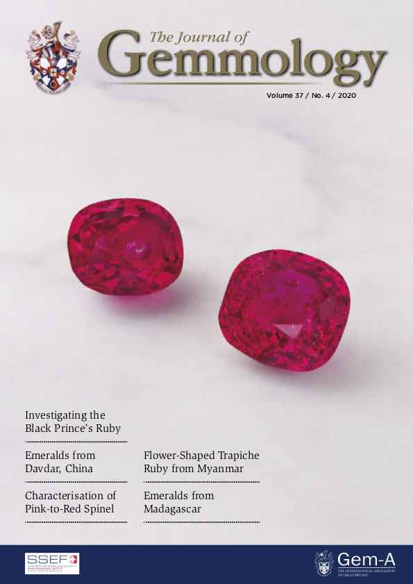
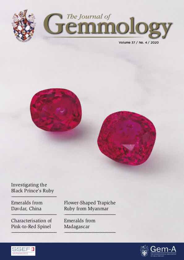
Characterisation of pink-to-red spinel from four important localities
Journal Article
Chankhanatha, C., Amphon, R., Rehman, H.U. and Shen, A.H. (2020) Characterisation of pink-to-red spinel from four important localities. Journal of Gemmology, Vol. 37, No. 4, pp. 393–403.
Record No. 6169  |  ISBN/ISSN: 221252  |  DOI: https://doi.org/10.15506/JoG.2020.37.4.393  |  Sinkankas/Born No.: NIS/NIB  |  RWHL
Most pink-to-red spinel that is currently available on the global gem market originates from moderate– to high–grade metamorphic rocks. For this study, we characterised 60 such spinels from Myanmar, Tajikistan, Tanzania and Vietnam. They ranged from pink to red with orange or blue modifiers due to combinations of the chromophores Cr³⁺, V³⁺ and Fe²⁺. The most common mineral inclusions in the samples from Myanmar, Vietnam and Tanzania were apatite crystals and graphite platelets, and we also observed zircon in the spinels from Tanzania. Octahedral negative crystals and partially healed fractures were present in samples from all four origins, while dislocations/needles were found only in those from Tanzania and Vietnam. Linear discriminant analysis of chemical data obtained by LA–ICP–MS was performed using seven elements (Ti, V, Cr, Fe, Zn, Ga and Sn) and provided potential criteria for separating spinels from these four important localities.

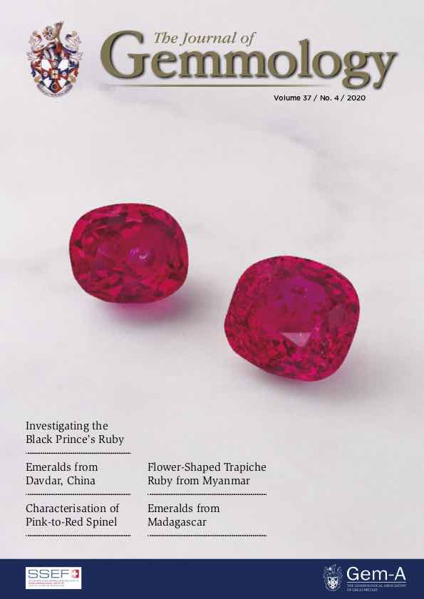
A study of emeralds from Davdar, North-Western China
Journal Article
Cui, D., Liao, Z., Qi, L., Zhong, Q. and Zhou, Z. (2020) A study of emeralds from Davdar, North-Western China. Journal of Gemmology, Vol. 37, No. 4, pp. 374–392.
Record No. 6168  |  ISBN/ISSN: 221252  |  Sinkankas/Born No.: NIS/NIB  |  RWHL
At the Davdar mine in Xinjiang, north-western China, emeralds are hosted mainly by carbonate, quartz–carbonate and quartz veins cutting metasedimentary rocks, and are associated with minerals such as hematite, dolomite, quartz, orthoclase and albite. Sixteen rough emeralds obtained during the authors’ visit to the mining area in 2019 were studied by standard gemmological techniques and various spectroscopic methods (FTIR, Raman, UV–Vis–NIR and EPR), as well as LA-ICP-MS chemical analysis. The analysed samples were mostly coloured by Cr, and showed a wide range of Fe, V, Mg and alkali contents, along with relatively low Cs, Rb and Sc. UV–Vis–NIR spectra showed features at 370 nm (Fe³⁺), 430 nm (Cr³⁺ with contributions from V³⁺ and possibly Fe³⁺), 580–630 nm (Cr³⁺ and V³⁺), 638 and 683 nm (Cr³⁺), and 850 nm (Fe²⁺ and possibly Fe²⁺–Fe³⁺ interactions). In addition, the more V-rich emeralds displayed a distinct V³⁺ absorption band at about 385–395 nm. Notably, the chemical composition of Davdar emeralds shows significant overlap with those from Panjshir, Afghanistan.

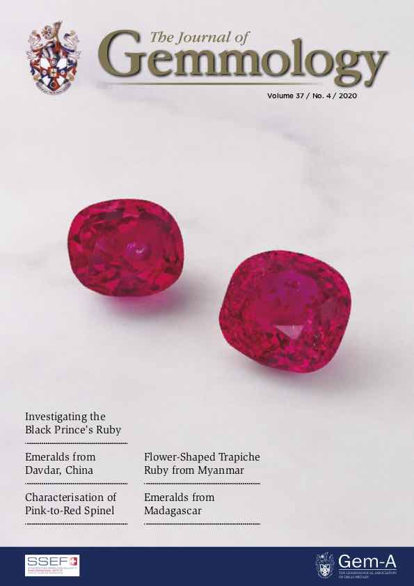
Tourmaline with cassiterite inclusions
Journal Article
Delaunay, A., Fritsch, E. and Hennebois, U. (2020) Tourmaline with cassiterite inclusions. Journal of Gemmology, Vol. 37, No. 4, pp. 349–350.
Record No. 6167  |  ISBN/ISSN: 221252  |  Sinkankas/Born No.: NIS/NIB  |  RWHL
Tourmaline is well known for its range of colours, but not so much for its variety of inclusions. Most of the time it simply contains elongated filamentary two-phase fluid inclusions referred to as ‘trichites’. While cataloguing some older samples in the collection of the French Gemmological Laboratory, we came across a tourmaline of unknown geographical origin that contained some unusual octahedral inclusions (Figure 8).

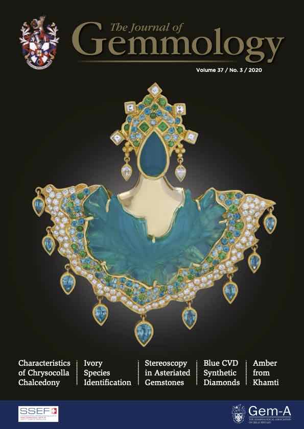
Vesuvianite (Idocrase) and apatite inclusions in ruby Identifiable by infrared spectroscopy
Journal Article
Smith, C.P. and Zellagui, R. (2020) Vesuvianite (Idocrase) and apatite inclusions in ruby Identifiable by infrared spectroscopy. Journal of Gemmology, Vol. 37, No. 4, pp. 346–348.
Record No. 6166  |  ISBN/ISSN: 221252  |  Sinkankas/Born No.: NIS/NIB  |  RWHL
Recently, two rubies weighing 1.39 and 2.19 ct (see the cover of this issue) were submitted to the American Gemological Laboratories (AGL) separately for testing, and they provided an opportunity to expand the range of inclusions identifiable in corundum using infrared spectroscopy. Routine origin testing indicated that both samples originated from Myanmar (Burma).

Gem Notes: A rare bicoloured spinel

Journal Article
Buathong, A. and Narudeesombat, N. (2020) Gem Notes: A rare bicoloured spinel. Journal of Gemmology, Vol. 37, No. 3, pp. 250–252.
Record No. 6128  |  ISBN/ISSN: 221252  |  Sinkankas/Born No.: NIS/NIB  |  RWHL
Buathong and Narudeesombat describe a rare natural spinel with distinct purple and greenish blue zones, investigating its identity, colour mechanisms, chemical composition, and treatment status. Submitted to the GIT Gem Testing Laboratory, the faceted specimen weighed 0.57 ct and measured 7.02 × 3.32 × 2.27 mm. It was singly refractive, with a refractive index of 1.718 and hydrostatic specific gravity of 3.60, consistent with spinel. Microscopy revealed oriented arrays of short to elongated iridescent silk and minute particles in the purple zone, while the greenish blue portion contained no observed inclusions. Both zones were inert under long- and short-wave ultraviolet radiation. Ultraviolet–visible–near-infrared spectroscopy showed absorptions at 371, 386, 458, and 555 nm in the purple zone. The greenish blue zone displayed corresponding features together with additional broad bands at approximately 656 and 917 nm. These absorptions were interpreted in terms of ferrous iron, ferric iron, and intervalence charge transfer between them. In the purple region, iron-related absorption leaves a blue-violet transmission window, while decreasing absorption towards the red contributes to the observed purple colour. Stronger short-wavelength absorption and the broad intervalence charge-transfer band near 656 nm instead produce a blue-green transmission window in the greenish blue region. The authors infer somewhat greater concentrations of ferrous and ferric iron in that zone, consistent with its higher measured total iron content. Semi-quantitative energy-dispersive X-ray fluorescence analysis identified magnesium and aluminium as major constituents, minor iron, silicon, and zinc, and traces of vanadium, chromium, and gallium. Total iron, expressed as Fe₂O₃, was 2.56 wt.% in the purple portion and 2.77 wt.% in the greenish blue portion; zinc oxide was 1.01 and 0.69 wt.%, respectively. Photoluminescence spectra obtained with 532 nm laser excitation showed a closely matching series of sharp chromium-related emission centres in both zones. These features were consistent with chromium substituting for aluminium in octahedral sites, and their sharpness supported identification as unheated material. The combination of characteristic inclusions, significant zinc content, physical properties, and photoluminescence behaviour established the specimen as natural, unheated spinel. The report documents how chemical differences and associated iron absorption can produce contrasting colours within a gem species in which a wide overall colour range is common but bicoloured specimens are scarce.

Gemmological and spectroscopic characteristics of different varieties of amber from the Hukawng Valley, Myanmar
Journal Article
Jiang, X., Zhang, Z., Wang, Y. and Kong, F. (2020) Gemmological and spectroscopic characteristics of different varieties of amber from the Hukawng Valley, Myanmar. Journal of Gemmology, Vol. 37, No. 2, pp. 144–162.
Record No. 6097  |  ISBN/ISSN: 221252  |  DOI: https://doi.org/10.15506/JoG.2020.37.2.144  |  Sinkankas/Born No.: NIS/NIB  |  RWHL
With its broad range of varieties, Burmese amber may show various optical effects, including ‘oil gloss’ (a red or green surface fluorescence effect) and phosphorescence phenomena. Samples representing golden, brown, red (‘blood’), ‘beeswax’ and ‘root’ ambers were characterised, as well as those showing red or green ‘oil gloss’; many of these also displayed phosphorescence. Common internal features seen with optical microscopy include reddish brown dot-like inclusions in brown amber and flow-banded concentrations of bubbles in ‘beeswax’ and ‘root’ ambers (the latter displaying patterns comprised of opaque light- and dark-coloured layers). SEM imagery revealed various scaly or layered structures on freshly broken surfaces, micro-bubbles in some samples and distinc- tive hollow micro-channels in ‘root’ amber. Fluorescence spectra recorded luminescence centres at 432 and 470 nm in all samples, and additionally at 650 nm (± 625 nm) in red ‘oil gloss’ amber. The ‘oil gloss’ appearance is caused by a mixture of surface fluorescence and the amber’s body colour. Phosphorescence spectroscopy showed that the effect was strongest at 525 nm, and the phosphores- cence lifetime measured at this emission wavelength ranged from 0.134 to 1.396 seconds.

Blue sapphire with partial red surface diffusion
Journal Article
Williams, B., Williams, C. and Carmona, C.I. (2020) Blue sapphire with partial red surface diffusion. Journal of Gemmology, Vol. 37, No. 1, pp. 23–24.
Record No. 6090  |  ISBN/ISSN: 221252  |  Sinkankas/Born No.: NIS/NIB  |  RWHL
A 1.94 ct cushion-cut blue sapphire was submitted for confirmation of treatment and identified as corundum using a GemmoRaman-532SG spectrometer. The stone exhibited a pale violetish blue body colour and deep reddish pink surface-related colouration restricted to the table and a small area along the pavilion. Microscopic observation revealed fingerprint inclusions, confirming the natural origin of the sapphire. Although the unusual colour distribution initially suggested a coating treatment on selected surfaces, careful microscopic examination showed that the pink colouration diffused slightly below the surface. Energy-dispersive X-ray fluorescence (EDXRF) spectroscopy indicated significant chromium (Cr), and the pink areas exhibited red fluorescence under long-wave ultraviolet excitation, unlike red-coated stones previously examined by the authors. These observations were consistent with chromium-diffused corundum. Infrared spectral features recorded using a GemmoFtir spectrometer confirmed that the sapphire had been heat treated, suggesting that red surface diffusion had been applied and that most of the diffused layer had subsequently been removed by polishing. However, the reason for retaining the diffusion treatment on only some surface areas remained unclear, given the resulting unnatural appearance. The case documents a natural blue sapphire with partial red surface diffusion and highlights the identification of this treatment through microscopic examination, EDXRF spectroscopy, ultraviolet fluorescence, and infrared spectroscopy. The authors also suggest that previously treated stones may be polished to remove near-surface treatments as demand for pale-coloured gemstones increases, potentially leading to more frequent encounters with such material.

Titanite (sphene) inclusions in ruby identified by infrared spectroscopy
Journal Article
Smith, C.P., Hartley, A. and Zellagui, R. (2020) Titanite (sphene) inclusions in ruby identified by infrared spectroscopy. Journal of Gemmology, Vol. 37, No. 1, pp. 11–12.
Record No. 6089  |  ISBN/ISSN: 221252  |  Sinkankas/Born No.: NIS/NIB  |  RWHL
A 7.79 ct unheated natural ruby submitted to the American Gemological Laboratories (AGL) contained numerous small, transparent, light yellow crystals, in addition to concentrations of rutile silk and a few partially healed fissures. Raman analysis of several crystals identified them as titanite (sphene). The mid-infrared spectrum of the ruby revealed an unusual absorption band centred at approximately 3482 cm⁻¹. Based on previous experience, the authors considered that this band was not intrinsically related to corundum and compared it with the spectrum of titanite, which also exhibits a dominant absorption band in this region. Overlaying the two spectra supported the interpretation that the 3482 cm⁻¹ absorption band recorded in the ruby was related to the dominant absorption in titanite. Mid-infrared spectroscopy has previously been used to identify mineral inclusions in gem corundum, including aluminium hydroxides such as diaspore and boehmite, calcite, weathering minerals such as kaolinite and goethite, micas and other minerals. Because corundum (Al₂O₃) is an anhydrous mineral, the mid-infrared spectral region between 5000 and 1800 cm⁻¹ can be useful for identifying mineral inclusions and determining the heated or unheated condition of ruby and sapphire.

G&G Microworld: Elongated rutile crystal in blue sapphire from Afghanistan
Journal Article
Tanawansombat, T. (2020) G&G Microworld: Elongated rutile crystal in blue sapphire from Afghanistan. Gems & Gemology, Vol. 56, No. 4, Winter, p. 531.
Record No. 4511  |  ISBN/ISSN: 0016626X  |  Sinkankas/Born No.: NIS/NIB  |  RWHL
Rutile, titanium dioxide (TiO₂), is a tetragonal mineral that generally occurs in corundum as silk (exsolution-formed needles), dust-like exsolution particles, or macroscopic crystal inclusions. Rutile crystals can have orange to deep red-brown coloration, and the crystal shapes can be rounded or euhedral. The author recently examined a 4.24 ct faceted transparent blue sapphire from Afghanistan. The stone contained dusty flake-like inclusions, naturally healed fractures, lathe-like inclusions, twinning, rounded colorless crystals identified as apatite (RRUFF R040098), and an unusual long prismatic orange crystal identified as rutile (RRUFF R040049). This rutile crystal shape is not typical for corundum, particularly in blue sapphire.

G&G Microworld: Zircon in jadeite jade
Journal Article
Jones, E. (2020) G&G Microworld: Zircon in jadeite jade. Gems & Gemology, Vol. 56, No. 4, Winter, pp. 531–532.
Record No. 4510  |  ISBN/ISSN: 0016626X  |  Sinkankas/Born No.: NIS/NIB  |  RWHL
One translucent green bangle bracelet recently examined by the author contained an interesting inclusion. The material was identified as dyed and polymer-impregnated jadeite jade using standard gemological testing and advanced spectroscopy, including UV-Vis, Raman, and infrared. This material would be classified as C-type jade in the trade. Microscopic examination revealed a surprisingly well-formed transparent brownish crystal inclusion. Raman analysis of the doubly terminated tetragonal prism revealed it to be “high” zircon (ZrSiO₄). To confirm this, the full width at half maximum (FWHM) of the internal stretching mode at 1008 cm⁻¹ was calculated as 6.05 cm⁻¹, indicating a high level of crystallinity, as this band is known to broaden as metamictization occurs (L. Nasdala et al., “Metamictization and U-PB isotopic discordance in single zircons: A combined Raman microprobe and SHRIMP ion probe study,” Mineralogy and Petrology, Vol. 62, 1998, pp. 1–27). Although interstitial zircon grains are not uncommon in jadeites, such a euhedral crystal is rarely observed and is especially unexpected considering the various treatments its host had undergone.

G&G Microworld: Uncommon inclusion of lazurite in ruby from Mogok, Myanmar
Journal Article
Aung, T. (2020) G&G Microworld: Uncommon inclusion of lazurite in ruby from Mogok, Myanmar. Gems & Gemology, Vol. 56, No. 4, Winter, p. 529.
Record No. 4509  |  ISBN/ISSN: 0016626X  |  Sinkankas/Born No.: NIS/NIB  |  RWHL
A parcel of small rubies was received for identification at the S Gemmological Institute (SGI) gem lab in Yangon, Myanmar. Based on chemical, spectroscopic, and standard gemological testing, they were determined to originate from the Mogok Stone Tract in Myanmar. One faceted ruby contained a small blue crystal inclusion with what appeared to be a pyritohedron shape but an uncommonly seen bright blue color (figure 9). This inclusion was identified by Raman spectroscopy as lazurite. A blue lazurite inclusion was previously reported to have been found in a 5.09 ct Burmese ruby and a 1.40 ct ruby from Namya, Myanmar (Spring 2012 Lab Notes, pp. 51–52). Also, blue lazurite mineral has previously been detected in 1.02 ct light gray Burmese spinel (Spring 2019 Micro-World, pp. 112–113). According to the literature and our knowledge of inclusions in rubies from Mogok, this blue mineral is rare as an inclusion in Burmese rubies.

G&G Microworld: Inclusions in Kenyan rubies from the John Saul mine
Journal Article
Khowpong, C. (2020) G&G Microworld: Inclusions in Kenyan rubies from the John Saul mine. Gems & Gemology, Vol. 56, No. 4, Winter, pp. 528–529.
Record No. 4508  |  ISBN/ISSN: 0016626X  |  Sinkankas/Born No.: NIS/NIB  |  RWHL
Rubies from the John Saul mine have been available in the market since the 1970s. The gems are found in a pegmatite-like vein that intrudes an ultrabasic body within the country rock (Winter 1999 Gem News, pp. 213–215). While the mine has produced a large quantity of ruby with fine color, only a small part of the production is fine quality and suitable for faceting, with most of the samples being cut into cabochons. Production has declined, and the material is seen less nowadays. The rubies have an inclusion scene that typically includes short, iridescent needles associated with dense clouds of particles, often in bands (figure 7). Negative crystals surrounded by a plane of thin films are also typical (figure 8). These are very similar to the inclusion scenes in rubies from Mogok (Myanmar) and other marble-hosted deposits. The fact that the color and fluorescence, which is dictated by the trace element chemistry, are also similar to marble-related deposits makes separating them from Kenyan rubies even more challenging. This shows that similar inclusion scenes can form in very different geological environments and that the internal world of gemstones is not always a conclusive indicator of geographic origin.

Staurolite in a Mozambique ruby

Journal Article
Ahline, N. (2020) Staurolite in a Mozambique ruby. Gems & Gemology, Vol. 56, No. 3, Fall, pp. 434–435.
Record No. 4422  |  ISBN/ISSN: 0016626X  |  Sinkankas/Born No.: NIS/NIB  |  RWHL
Discovery of staurolite in Mozambique ruby.

Lab Notes: Negative crystal containing a mobile CO₂ bubble in blue sapphire heated with pressure
Journal Article
Ng-Pooresatien, N. (2020) Lab Notes: Negative crystal containing a mobile CO₂ bubble in blue sapphire heated with pressure. Gems & Gemology, Vol. 56, No. 2, Summer, pp. 287–288.
Record No. 4333  |  ISBN/ISSN: 0016626X  |  Sinkankas/Born No.: NIS/NIB  |  RWHL
Among the common inclusions seen in almost every mineral are primary cavities, or negative crystals. Negative crystals in sapphire are usually filled with liquid and gaseous carbon dioxide (CO₂) and may also contain solids such as diaspore or graphite. At GIA’s Bangkok laboratory, the author recently examined a 6.41 ct transparent blue faceted sapphire. It had a refractive index of 1.760–1.768, and its fluorescence reaction was inert under long- and short-wave UV radiation. Fourier-transform infrared (FTIR) spectroscopy is a useful technique to determine heat treatment in corundum. The sample’s FTIR spectrum showed a broad band centered around 3047 cm⁻¹ that has been reported in sapphires treated with heat and pressure. Microscopic observation revealed altered growth tubes, dissolved particles, melted crystals with altered fingerprints, and small birefringent crystals in a low-relief partially healed fissure—proof of high-temperature heat treatment with pressure. When the sapphire was gently heated by the microscope bulb, the gas bubble became smaller and disappeared as the gas and liquid homogenized. The presence of CO₂ fluid inclusions in negative crystals is important evidence of natural origin and the absence of thermal enhancement. Interestingly, this sample exhibited a rounded bubble in a negative crystal that moved and was still observed when gently heated with a hot point. Raman spectroscopy confirmed the bubble as carbon dioxide (CO₂). To our knowledge, a mobile CO₂ bubble in a negative crystal has not been reported in a sapphire heated with pressure.

G&G Micro-World: Unusually shaped rutile crystal inclusions in Russian emerald
Journal Article
Khowpong, C. (2020) G&G Micro-World: Unusually shaped rutile crystal inclusions in Russian emerald. Gems & Gemology, Vol. 56, No. 2, Summer, p. 294.
Record No. 4332  |  ISBN/ISSN: 0016626X  |  Sinkankas/Born No.: NIS/NIB  |  RWHL
Russian emeralds have been mined since the mid-nineteenth century near Yekaterinburg in the Ural Mountains. They are classified as schist-related emeralds, meaning they formed in the reaction zone of a pegmatite intruding into schists. Similar emeralds are found in Zambia, Brazil, and Ethiopia. All of these deposits can show a similar inclusion scene (S. Saeseaw et al., “Geographic origin determination of emerald,” Winter 2019 G&G, pp. 614–646). A 1.489 ct Russian emerald crystal collected by GIA at the Mariinsky Priisk mine showed several interesting included crystals. The crystals appeared as long yellowish brown rods (figure 6). In some cases, they appeared as a group of disordered coarse needles (figure 7). They were identified by Raman spectroscopy as rutile. Although rutile crystal inclusions are common in schist-related emeralds, this crystal morphology is unusual and has not previously been mentioned in the literature. Therefore, this inclusion might be unique and different from the other schist-related emerald sources. These unusual patterns could potentially be an indicator of Russian origin.

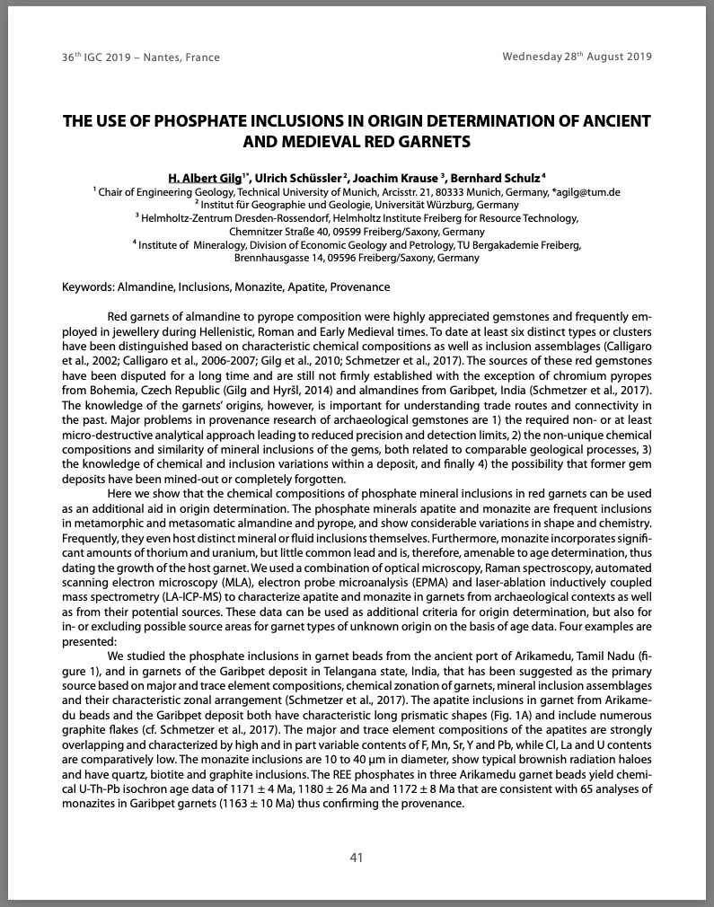
The use of phosphate inclusions: Origin determination of ancient and medieval red garnets
Conference Paper
Gilg, H.A., Schüssler, U., Krause, J. and Schulz, B. (2019) The use of phosphate inclusions: Origin determination of ancient and medieval red garnets. 36th International Gemmological Conference, Nantes, France pp. 41–43.
Record No. 26810  |  Sinkankas/Born No.: NIS/NIB  |  RWHL
Red garnets of almandine to pyrope composition were highly valued gemstones and commonly used in jewelry throughout the Hellenistic, Roman, and Early Medieval periods. At least six distinct types or compositional clusters have been identified based on characteristic chemical signatures and inclusion assemblages (Calligaro et al., 2002; Calligaro et al., 2006–2007; Gilg et al., 2010; Schmetzer et al., 2017). Despite their historical prevalence, the precise sources of most red garnets remain uncertain, with the exception of chromium-rich pyropes from Bohemia, Czech Republic (Gilg and Hyršl, 2014), and almandines from Garibpet, India (Schmetzer et al., 2017). Determining the origin of these garnets is vital for reconstructing ancient trade routes and patterns of cultural exchange. However, several challenges complicate provenance research: (1) the need for non-destructive or minimally invasive analytical methods, which limit precision and sensitivity; (2) the non-unique chemical and inclusion features caused by similar geological formation processes; (3) insufficient knowledge of compositional and inclusion variability within individual deposits; and (4) the possibility that some ancient sources have been exhausted or lost from historical record.

Infrared spectra and inclusions of emeralds from Pakistan
巴基斯坦祖母绿的红外光谱和包裹体特征研究
Journal Article
Guo, H. and Yu, X. (2019) Infrared spectra and inclusions of emeralds from Pakistan. [巴基斯坦祖母绿的红外光谱和包裹体特征研究] Acta Petrologica et Mineralogica [岩石矿物学杂志], Vol. 38, No. 5, pp. 724–732.
Record No. 17171  |  Sinkankas/Born No.: NIS/NIB  |  Language: in Chinese with English abstract  |  RWHL
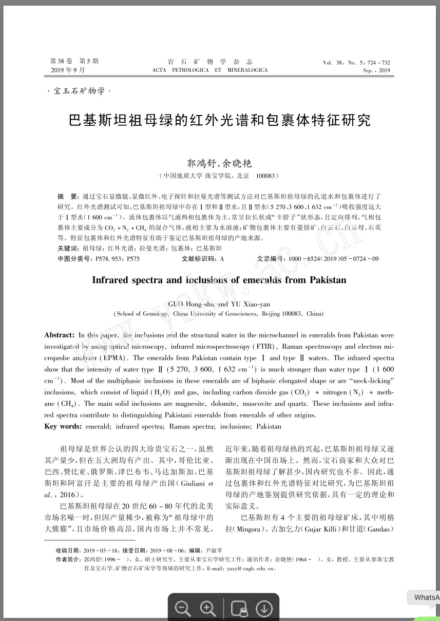
In this paper, the inclusions and the structural water in the microchannel in emeralds from Pakistan were investigated by using optical microscopy, infrared microspectroscopy (FTIR), Raman spectroscopy, and electron microprobe analyzer (EPMA). The emeralds from Pakistan contain type I and type II waters. The infrared spectra show that the intensity of water type II (5270, 3600, 1632 cm⁻¹) is much stronger than water type I (1600 cm⁻¹). Most of the multiphasic inclusions in these emeralds are of biphasic elongated shape or are “neck-licking” inclusions, which consist of liquid (H₂O) and gas, including carbon dioxide gas (CO₂) + nitrogen (N₂) + methane (CH₄). The main solid inclusions are magnesite, dolomite, muscovite, and quartz. These inclusions and infrared spectra contribute to distinguishing Pakistani emeralds from emeralds of other origins.

A study of mineral inclusions in emeralds from Malipo, Yunnan Province, China
Journal Article
Jiang, X., Yu, X., Guo, B. and Xu, C. (2019) A study of mineral inclusions in emeralds from Malipo, Yunnan Province, China. Acta Petrologica Et Mineralogica, Vol. 38, No. 2, pp. 279–286.
Record No. 17168  |  Sinkankas/Born No.: NIS/NIB  |  Language: in Chinese with English abstract  |  RWHL
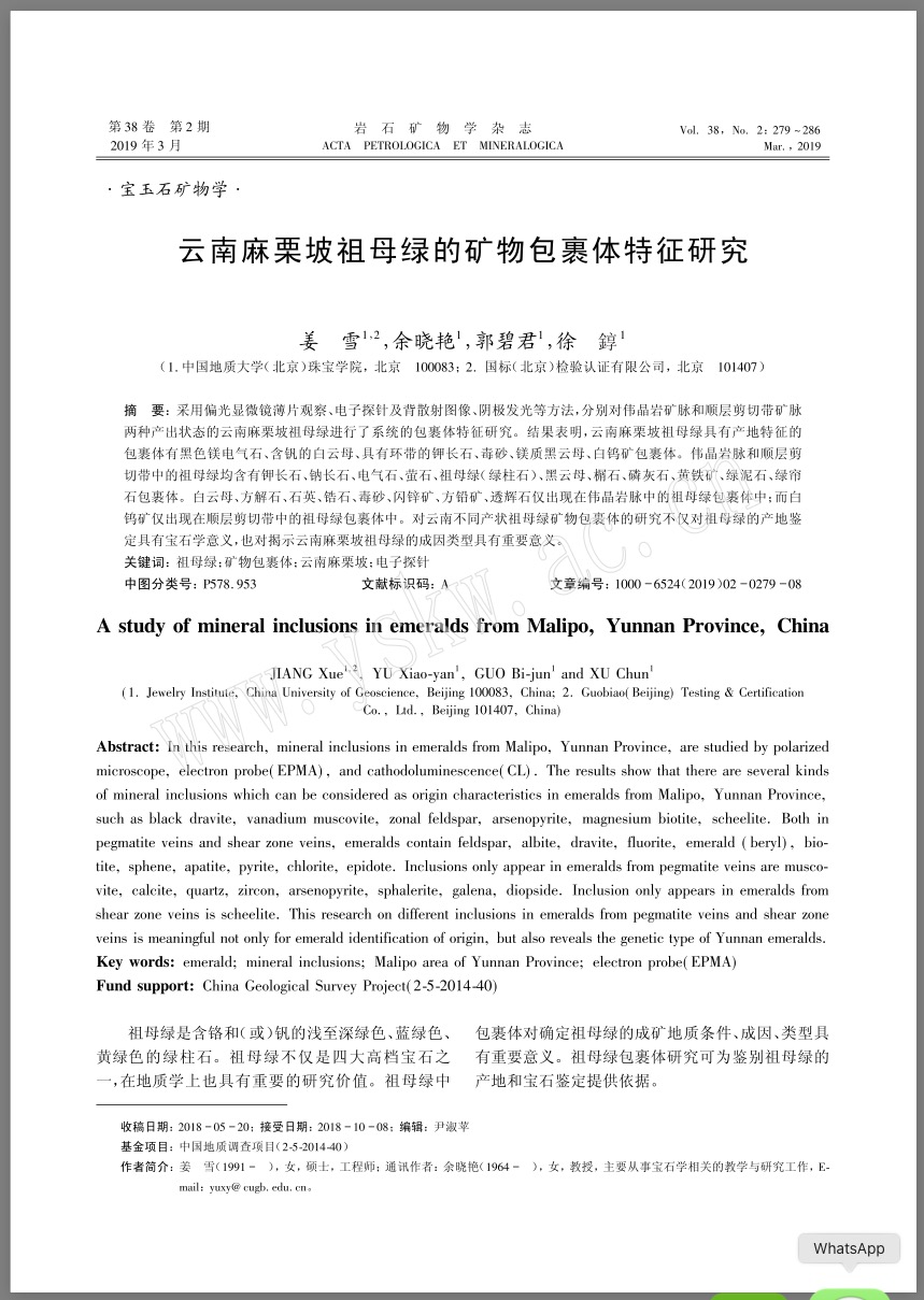
In this research, mineral inclusions in emeralds from Malipo, Yunnan Province, are studied by polarized microscope, electron probe(EPMA), and cathodoluminescence(CL). The results show that there are several kinds of mineral inclusions which can be considered as origin characteristics in emeralds from Malipo, Yunnan Province, such as black dravite, vanadium muscovite, zonal feldspar, arsenopyrite, magnesium biotite, scheelite. Both in pegmatite veins and shear zone veins, emeralds contain feldspar, albite, dravite, fluorite, emerald (beryl), biotite, sphene, apatite, pyrite, chlorite, epidote. Inclusions only appear in emeralds from pegmatite veins are muscovite, calcite, quartz, zircon, arsenopyrite, sphalerite, galena, diopside. Inclusion only appears in emeralds from shear zone veins is scheelite. This research on different inclusions in emeralds from pegmatite veins and shear zone veins is meaningful not only for emerald identification of origin, but also reveals the genetic type of Yunnan emeralds.

Orange-yellow inclusion in spinel from Man Sin, Myanmar

Journal Article
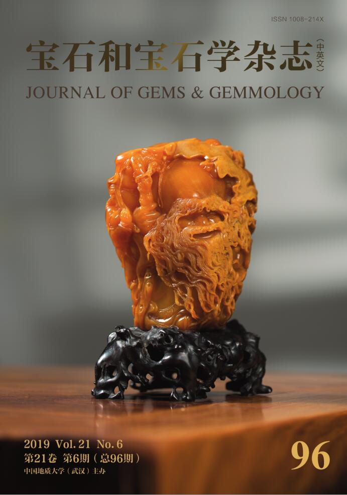
Zhai, S., Pei, J. and Huang, W. (2019) Orange-yellow inclusion in spinel from Man Sin, Myanmar. Journal of Gems & Gemmology, Vol. 21, No. 6, pp. 24–30.
Record No. 16617  |  DOI: https://dx.doi.org/10.15964/j.cnki.027jgg.2019.06.003  |  Sinkankas/Born No.: NIS/NIB  |  Language: in Chinese with English abstract  |  RWHL
Providing the famous “Jedi” spinel, Man Sin (Myanmar) has gained more and more attention in these years. Gem microscope and Raman spectrometer were used to carefully test inclusions in spinels. The multi-phase inclusion containing orange-yellow fluid could help distinguish Man Sin spinels from the spinels of other localities. The orange-yellow fluid was found in octahedral negative crystals and on the surface of mineral inclusions, which could form the indicating multi-phase inclusions with H₂S (gaseous and liquid) and a variety of minerals. The Raman spectrum of the orange-yellow fluid shows four bands at 155, 220, 443, 475 cm⁻¹ which indicate that it is high-level natural sulphur solution and the liquid is probably CS₂. Sulphur indicates highly reducing conditions during the formation of spinels from Man Sin, Myanmar.

Exploring the Literature of Gemstone Inclusions

The literature of gemstone inclusions covers identification, origin determination, treatments, synthesis, mineral inclusions, fluid inclusions and other internal features of natural and synthetic gems.

For a curated selection of important sources, read Inclusions in Gemstones — Hyperion Literature Sources.

About Four Treasures

Four Treasures is Lotus Gemology's freely accessible gemological reference database. Use the full database to search by author, title or keyword and explore literature on gemology, mineralogy, jewelry and related fields. Where available, hyperlinks on titles may lead directly to the full publication.

Search the Complete Four Treasures Database