
Gemstone Inclusions Bibliography

Gemstone Inclusions Bibliography

Explore books, articles and historical sources on inclusions in gemstones in Four Treasures, the Lotus Gemology reference database. This bibliography brings together matching records from the collection, with publication details, abstracts and cover images where available.

Gemstone Inclusion References

The list below matches the terms inclusion or inclusions in the database's searchable fields. Some references discuss inclusions as part of a broader subject.
601–620 of 1,197 matching references

Study on gemmological characteristics and mineral components of Qinghai black nephrite
青海墨色软玉的宝石学特征及矿物组成研究
Thesis
Qin, Y. (2013) Study on gemmological characteristics and mineral components of Qinghai black nephrite. [青海墨色软玉的宝石学特征及矿物组成研究]., Beijing, China: China University of Geosciences (Beijing), 74 pp.
Record No. 7264  |  Sinkankas/Born No.: NIS/NIB  |  Language: in Chinese with English abstract  |  RWHL
At present, more and more black nephrite jade has appeared in both domestic and international markets, with much of it originating from newly discovered deposits in the Mo ore region of Qinghai. Using modern analytical techniques, the gemological and mineralogical characteristics of black nephrite from Qinghai have been systematically studied, and its genetic type has been inferred based on geological context. Polarizing microscope observations show that the main mineral in Qinghai black nephrite is tremolite, with numerous internal filling cracks and inclusions of graphite, magnetite, ilmenite, and other mineral impurities. Scanning electron microscopy reveals that its primary texture is columnar, with microstructural features including fibrous interwoven textures and scaly metacrystic structures. Infrared spectroscopy of representative samples shows absorption peaks corresponding to tremolite, with the peak positions unaffected by color variation. X-ray diffraction analysis confirms that Qinghai black nephrite closely matches the unit cell parameters of standard tremolite. Differential thermal analysis shows an endothermic effect during heating to 1000 °C, attributed to the loss of interlayer and structural water, with weight loss ranging from 1.315% to 5.058%. Electron probe analysis yields Mg²⁺/(Mg²⁺+Fe²⁺) ratios above 0.9, confirming classification within the tremolite series. The formation of color in the nephrite does not appear to be related to typical chromophoric elements such as Ti, Mn, Cr, or Fe, and no clear correlation was found between color and the substitution of Mg–Si, (Ca+Na+K)–Al, or (Ca+Na+K)–Fe in the crystal structure. Comparative analysis with black nephrite from Hotan and Xiuyan indicates that Qinghai black nephrite has slightly higher levels of calcium, magnesium, and silicon. Based on the regional geology of Qinghai, it is inferred that this nephrite formed through hydrothermal metasomatism of dolomitic marble, with graphite, magnetite, and other inclusions being incorporated into tremolite during the hydrothermal stage.

Gemological characteristics of Russian Gorlik-gol jasper and research on composition of melanocratic mineral
俄罗斯哥力-哥尔矿碧玉的宝石学特征及暗色斑点成分研究
Thesis
Zhang, S. (2013) Gemological characteristics of Russian Gorlik-gol jasper and research on composition of melanocratic mineral. [俄罗斯哥力-哥尔矿碧玉的宝石学特征及暗色斑点成分研究]., Beijing: China University of Geosciences
Record No. 7047  |  Sinkankas/Born No.: NIS/NIB  |  Language: in Chinese with English abstract  |  RWHL
Because the Ospa (No. 7) yu is still actively mined in Russia and the Gorlik-gol (No. 10) yu is widely available, Russian yu holds a significant share of the Chinese market. Given the confusion within China’s yu market and the absence of a precise classification for yu in the National Treasure Yu Stone standard, this study conducts a comprehensive analysis of yu from the Gorlik-gol area, focusing on gem-mineralogical characteristics, chemical and physical properties, and the composition of dark inclusions. Analytical techniques include polarized light microscopy, scanning electron microscopy (SEM), Fourier-transform infrared spectroscopy (FTIR), X-ray powder diffraction (XRD), and electron probe microanalysis (EPMA). Gorlik-gol yu exhibits colors ranging from green to dark green, a greasy to waxy luster, slight translucency to opacity, a refractive index of approximately 1.60, Mohs hardness between 5 and 6, an average density of 2.96 g/cm³, and irregular fracture surfaces. Microscopically, the dominant mineral is fibrous tremolite, displaying textures such as felted fibrous crystalloblastic, micro-fibrous metacrystic, fibrous radial, and nearly metacrystalline structures. SEM reveals interwoven fibrous structures, directional micro-fiber alignments, weakly oriented textures, microsheet formations, and burr-like features. XRD and EPMA confirm tremolite as the main mineral (>94%), with some samples containing actinolite as the primary phase (>63%). Secondary minerals include chlorite, iron chromite, Fe-Mg chromite, fluorapatite, and copper sulfide. Yu-like stones near the Gorlik-gol deposit are similar, with tremolite as the main phase and secondary minerals such as clinochlore, alumino-magnesian chromite, copper sulfide, and quartz. The dark-colored inclusions consist mainly of Fe-Mg chromite, iron chromite, and copper sulfide. Elevated FeO and MnO levels suggest that surrounding yu may be erosional derivatives of the Gorlik-gol source. Well-developed conjugated X-type shear joints due to late-stage tectonic compression have fragmented the chromite, suggesting a three-phase chromite formation history. The Gorlik-gol yu deposit is interpreted as a metamorphic hydrothermal deposit hosted in ultramafic ophiolitic rocks intersected by rodingitized veins.

Study on the gemmological and mineralogical characteristics of green nephrite in Hualian, Taiwan
台湾花莲碧玉宝石矿物学特征研究
Thesis
Liu, D. (2013) Study on the gemmological and mineralogical characteristics of green nephrite in Hualian, Taiwan. [台湾花莲碧玉宝石矿物学特征研究]., Beijing: China University of Geosciences
Record No. 6951  |  Sinkankas/Born No.: NIS/NIB  |  Language: in Chinese with English abstract  |  RWHL
China has a long history of jade culture, with nephrite taking first place among the “Four Famous Jades,” giving it a broad market and widespread popularity among consumers. In this article, testing methods including polarizing microscopy, scanning electron microscopy, electron microprobe analysis, X-ray powder diffraction (XRD), and Fourier transform infrared spectroscopy (FTIR) were used to investigate the gemological and mineralogical characteristics of green nephrite from Hualien, Taiwan. Scientific, comprehensive, and systematic research is of great significance for enhancing the scientific understanding and recognition of Hualien nephrite. Observations show that the color of green nephrite ranges from light green to dark green, with relatively pure but unevenly distributed coloration. Black metallic mineral inclusions are present. The nephrite exhibits a lipidic-silky luster and is translucent to opaque. Its refractive index is 1.61, with a relative density of 2.96 to 3.02 and a hardness between 6 and 7. The primary mineral is either tremolite or actinolite, while secondary minerals include grossular, chromite, and chlorite. Electron microprobe analysis shows that the main components are MgO, SiO₂, and CaO, with small amounts of FeO and trace levels of Cr₂O₃, MnO, K₂O, Na₂O, Al₂O₃, and TiO₂. Comparison of sample data suggests that the depth of green coloration is positively correlated with FeO content, and the presence of grossular enhances color brightness. XRD results confirm that the main mineral is tremolite or actinolite. Diffraction lines of the surrounding rock match serpentine standards and indicate limited tremolite, suggesting close growth and mineral transition at the contact zone, where alteration of the host rock has occurred. FTIR analysis shows that the OH bands are separated and shifted to lower wavenumbers, likely due to Fe substituting for Mg. The absence of stretching vibrations from adsorbed water and a lack of significant absorption bands indicate that water did not occupy internal or surface space during nephrite formation. Polarizing and scanning electron microscopy reveal two microstructures: felt-like metacrystic and parallel-fibrous metacrystic, with the overall structure being schistose, while the host rock exhibits a massive texture. Comparison with nephrite from other localities shows similar characteristics, making origin identification difficult without detailed analysis of gemological features and mineral composition. The presence of locality-specific secondary minerals may aid in distinguishing between nephrites from different sources. Green nephrite from Hualien, Taiwan, is prized for its vivid color, fine texture, and unique optical effects. High-quality specimens have significant economic value, and the rare green nephrite cat’s-eye variety holds the highest value of all.

Is Myanmar jadeitite of Jurassic age? A result from incompletely recrystallized inherited zircon
Journal Article
Yui, T.-F., Fukoyama, M., Iizuka, Y., Wu, C.-M., Wu, T.-W., Liou, J.G. and Grove, M. (2013) Is Myanmar jadeitite of Jurassic age? A result from incompletely recrystallized inherited zircon. Lithos, Vol. 160–161, pp. 268–282.
Record No. 5806  |  Sinkankas/Born No.: NIS/NIB  |  RWHL
Zircons from two Myanmar jadeitite samples were examined for texture, mineral inclusions, U–Pb dating, and trace element composition, revealing three distinct zircon types based on U–Pb isotopic characteristics. Type I zircons are inherited and yield an igneous protolith age of 160 ± 1 Ma; Type II zircons formed through metasomatic/hydrothermal processes and provide a minimum jadeitite formation age of 77 ± 3 Ma; and Type III zircons are incompletely recrystallized, with discordant and geologically meaningless U–Pb ages ranging from 153 to 105 Ma. These findings indicate that Myanmar jadeitites formed primarily by wholesale metasomatic replacement. Compared to Type I, Type II zircons display typical metasomatic/hydrothermal geochemical signatures, including low Th/U ratios (< 0.1), small Ce anomalies (Ce/Ce* < 5), and low total REE contents (40–115 ppm). Type III zircons exhibit intermediate characteristics. The study reveals that the resetting of trace elements and U–Pb isotopic systems in inherited zircons is asynchronous during jadeitite formation, and some Type I zircons with low Th/U and Ce anomalies likely reflect secondary modification. In extreme cases, incompletely recrystallized zircons may acquire hydrothermal-like geochemical signatures while retaining inherited U–Pb isotopic ages, leading to misinterpretation of jadeitite formation timing. The data support a Late Cretaceous formation age for Myanmar jadeitites, likely related to subduction processes predating the Paleocene India–Asia collision, and cast doubt on previously proposed Late Jurassic ages, which appear to have been derived from Type III zircons.

Jadeitite formed during subduction: In situ zircon geochronology constraints from two different tectonic events within the Guatemala Suture Zone
Journal Article
Flores, K.E., Martens, U.C., Harlow, G.E., Brueckner, H.K. and Pearson, N.J. (2013) Jadeitite formed during subduction: In situ zircon geochronology constraints from two different tectonic events within the Guatemala Suture Zone. Earth and Planetary Science Letters, Vol. 371–372, pp. 67–81.
Record No. 5805  |  Sinkankas/Born No.: NIS/NIB  |  RWHL
Jadeitite is a rare rock type associated with high-pressure–low-temperature blocks within serpentinite matrix mélanges. Models of formation involve precipitation from subduction-zone aqueous fluids veining the overlying mantle wedge (P-type), or metasomatism of igneous and/or sedimentary protoliths previously emplaced into the mélange (R-type). Age determinations of mélange lithologies provide constraints on the timing of “peak metamorphism” and subsequent exhumation. The timing of jadeitite formation, particularly in the rich source of the Guatemala Suture Zone (GSZ), is a controversial subject needing further attention. Over 80 in situ zircon crystals from three jadeitites and two mica–albite rocks from the North Motagua Mélange and one phengite jadeitite from the South Motagua Mélange of the GSZ were studied for age and trace-element determination. Most of these zircons are characterized by low Th/U ratios, depleted chondrite-normalized REE patterns relative to zircons from oceanic gabbros, and contain fluid and mineral inclusions that reflect the primary mineralogy (i.e., jadeite) and context (i.e., crystallization from an aqueous fluid) of the host rock, and thus formed during jadeitite crystallization. The SHRIMP-RG and LAM-ICP-MS U–Pb dates from zircon indicate that jadeitites and mica–albite rocks from the GSZ were formed through vein precipitation at 98–80 and 154–158 Ma, respectively. These data show (a) older ages that indicate jadeitite crystallization occurred 10–30 Ma before the preserved subduction-zone peak metamorphism (e.g., exhumed eclogite), and (b) a second group of ages slightly younger than, or similar to, exhumation ages given by Ar–Ar dates from micas. Similar relationships occur at other jadeitite occurrences, such as the Syum-Keu ultramafic complex in the Polar Urals (Russia) and the serpentinite mélanges of the Río San Juan complex (Dominican Republic). The data argue for formation of jadeitite within the mantle wedge during active subduction. Thus, jadeitite provides a record of fluid introduction into the mantle wedge during subduction rather than during exhumation.

Inclusions of black-green serpentine jade determined by Raman spectroscopy
Journal Article
Wang, Y.Y., Gan, F.X. and Zhao, H.X. (2013) Inclusions of black-green serpentine jade determined by Raman spectroscopy. Vibrational Spectroscopy, Vol. 66, May,, 2013/05/01/ pp. 19–23.
Record No. 5778  |  ISBN/ISSN: 9242031  |  DOI: https://doi.org/10.1016/j.vibspec.2013.01.008  |  Sinkankas/Born No.: NIS/NIB  |  RWHL
This work aims to determine the formation mechanism as well as the major mineral and inclusions of black-green serpentine jade by Raman spectroscopy. Scanning electron microscopy with energy-dispersive spectrometry was used to analyze the chemical composition of the inclusions and major mineral. The major mineral of black-green serpentine jade was antigorite, and the inclusions were actinolite, chlorite, calcite, quartz, magnetite, and goethite. Jade quality was preliminarily evaluated based on the area ratio of antigorite to the inclusions by optical microscopy. Formation mechanism of black-green serpentine jade was inferred based on the analysis of the inclusions, which demonstrated a new application of Raman spectroscopy in mineralogy.

A comparative overview of inclusions in spinel
Conference Paper
Boehm, E.W. (2013) A comparative overview of inclusions in spinel. International Gemmological Conference, Hanoi, Vietnam p. 88.
Record No. 4150  |  Sinkankas/Born No.: NIS/NIB  |  RWHL
Spinels contain many interesting inclusions that reveal secrets about their host rocks and sometimes also their geographic origin. The primary spinel producing countries are Burma (Myanmar), Sri Lanka, Madagascar, Tajikistan, Tanzania, and Vietnam. Spinel host rocks vary but may also have much in common, particularly those from metamorphosed dolomitic limestones (≥50% Mg replacing Ca) commonly referred to as marble. During metamorphism, calcic limestones become Magnesium rich which combines with silica to form spinel and silicates such as diopside, epidote, forsterite, garnet, zircon, etc. These same minerals may become characteristic inclusions within spinel. Other minerals that may be found in spinel include apatite, böhmite, diaspore, graphite, hercynite, högbomite, ilmenite, magnetite, various micas, rutile, sphene, and uraninite. These inclusions may be euhedral, subhedral, or corroded. The most diagnostic and commonly seen inclusions in natural spinels are octahedral negative crystals (Figure 1) that are often aligned in rows or multiple rows within their host. These negative crystals may also be seen containing white dolomite, yellow phlogopite, or black hercynite. Some inclusions or combinations of inclusions may be considered diagnostic for particular geographic origins of spinel. Transparent to white rounded apatite crystals together with attached black hexagonal crystals of ilmenite or graphite form a diagnostic inclusion scene in spinels from Burma (Myanmar). Högbomite on twinning planes is unique to spinels from Morogoro, Tanzania. Sri Lankan spinels often have an assemblage of apatite (Figure 2), diopside, phlogopite, rutile, sphene, uraninite, and zircon. Synthetic spinels are most often produced by the flame fusion process, which provides several diagnostic inclusions. Flux synthetic spinels may have typical flux inclusions but are often faceted into flawless gems, which require additional testing for conclusive identification. Inclusions in spinels are considered more useful for identifying their host than for origin determination, however, in some cases, diagnostic inclusions are evident.

Vibrational spectra of black species of Hetian nephrite (tremolite jade) and its color genesis
新疆和田黑色透闪石质软玉振动光谱特征及颜色成因
Journal Article
Yang, X., Shi, G. and Liu, Y. (2012) Vibrational spectra of black species of Hetian nephrite (tremolite jade) and its color genesis. [新疆和田黑色透闪石质软玉振动光谱特征及颜色成因] Spectroscopy and Spectral Analysis [光谱学与光谱分析], Vol. 32, No. 3, March, pp. 681–685.
Record No. 7301  |  Sinkankas/Born No.: NIS/NIB  |  Language: in Chinese with English abstract  |  RWHL
Black nephrite is a very uncommon jade type currently found only at Karakashi. In order to study the spectral characteristics and color-genetic mechanism of the black nephrite, samples collected from Karakashi were studied using EMPA, IR, Raman, and XRD. The results are as follows: nephrite with different colors has nearly the same major chemical compositions, while the black ones contain higher carbon. Nephrite with different colors has a similar vibration spectrum, while the black ones show the characteristic peak of graphite. It is speculated that the color of black nephrite is not only related to actinolite but also related to the graphite inclusions. Obviously, Raman spectra are a very important nondestructive method to identify the color genesis and the origin of the black nephrite.

Study on the Chuncheon nephrite deposit, Korea–Analysis on the mineralization and genesis
韩国春川软玉矿床研究–成矿作用及成因分析
Thesis
Pei, X. (2012) Study on the Chuncheon nephrite deposit, Korea–Analysis on the mineralization and genesis. [韩国春川软玉矿床研究–成矿作用及成因分析]., Beijing, China: China University of Geosciences (Beijing), 75 pp.
Record No. 7262  |  Sinkankas/Born No.: NIS/NIB  |  Language: in Chinese with English abstract  |  RWHL
The Chuncheon nephrite deposit is the largest in South Korea with an estimated reserve of 300,000 tons. Located in mid-eastern South Korea, the deposit is in the form of lenses in the contact zone between dolomitic marble and amphibole schist in the upper part of a biotite schist. To date, the origin, process of formation, and age of the Chuncheon nephrite deposit remain unclear, which hinders our further understanding of the formation of nephrite. This study involved petrography, chemical analyses, and nephrite age determinations using optical mineralogy, electron microprobe-analyses (EMPA), X-ray fluorescence spectroscopy (XRF), and SHRIMP (Sensitive High-Resolution Ion MicroProbe) analyses of zircon. Petrographic studies indicated that the Chuncheon nephrite consists mainly of tremolite. The low contents of Cr (2.15–4.74 ppm) and Ni (2.95–4.90 ppm) are typical of dolomite-related nephrites and lower than in serpentine-related nephrites. The total REE contents are <18.5 ppm. The REE chondrite-normalized patterns display right-leaning LREE and almost flat HREE patterns, which are consistent with the REE model of dolomitic marble. The δ¹⁸O isotope compositions of the zircons (6.34‰–12.5‰) from the nephrite overlap those of the Chuncheon dolomite (2.4‰–18.2‰), indicating clearly that dolomite played an important role in the formation of the Chuncheon nephrite. The petrography and geochemistry suggest that it had a metasomatic origin involving the reactions dolomitic marble → diopside, diopside → tremolite, and tremolite → chlorite. The δD and δ¹⁸O isotope compositions of our specimens of nephrite (δD = −95.3‰ to −78.3‰ and −118‰ to −105‰; ¹⁸O = −9.9‰ to −7.9‰) and the hydrothermal fluids in equilibrium with the nephrite (δD = −73.6‰ to −56.6‰ and −96.3‰ to −83.3‰, 330–450 °C; δ¹⁸O = −9.41‰ to −6.38‰, 330–430 °C) suggest that the hydrothermal fluids were predominantly meteoric water. The zircons contain almost no mineral inclusions, which is quite different from zircons in other nephrite deposits. The variable SHRIMP U–Pb ages of the zircons (908.9 ± 4.8 to 459.1 ± 1.9 Ma (n = 7), 436.3 ± 16.9 to 359.5 ± 6.4 Ma (n = 5), and 249.9 ± 5.2 to 79.4 ± 1.9 Ma (n = 5)) in the Chuncheon nephrite may record the ages of a regional multi-stage metamorphism. Due to the generally low temperature of formation of the nephrite, we speculate that the ages of zircons only record the two relatively high-temperature initial stages of nephrite formation (dolomitic marble → diopside, and diopside → coarse-grained tremolite) and failed to record the age of the lowest-temperature third stage of high-quality nephrite formation (coarse-grained tremolite → fine-grained tremolite). It has generally been considered that dolomite-related nephrite forms at the contacts between granite/granodiorite and dolomitic marble, whereas serpentinite-related nephrite forms at the contacts between serpentinite and silica-rich rocks. However, the genesis of the Chuncheon nephrite deposit does not fit with either of these well-known types. Thus, we developed a new model for the Chuncheon nephrite in which heat was provided by regional multi-stage metamorphism, meteoric water dominated the nephrite-forming fluids, and dolomitic marble and biotite schist provided the material for the nephrite.

Gemological characteristics of nephrite jade from Huanren, Liaoning
辽宁桓仁软玉的宝石学特征研究
Journal Article
Lin, W. (2012) Gemological characteristics of nephrite jade from Huanren, Liaoning. [辽宁桓仁软玉的宝石学特征研究] Rock and Mineral Analysis [岩矿测试], Vol. 31, No. 5, October, pp. 794–797.
Record No. 7249  |  Sinkankas/Born No.: NIS/NIB  |  Language: in Chinese with English abstract  |  RWHL
By using routine gemological testing, thin section observation under a microscope, Fourier Transform Infrared (FTIR) Spectrometric analysis, and the chemical analysis method, a systematic study of nephrite jade from Huanren has been conducted for minerals, chemical compositions, and gemological characteristics. The results indicate a refraction index of 1.60, specific density of 3.08, and Mohs hardness of about 6.5. Thin section observation by a polarizing microscope shows that the main mineral composition is epidote and hornblende, and the impurity minerals include diopside, apatite, carbonate minerals, and serpentine, among others. The main textures of the jade are inequigranular blastic, foliated pilotaxitic, and metasomatic. The specific texture characteristics of the nephrite are responsible for its poor transparency, uneven color distribution, and the inclusions of black spots and green agglomerate. The features of the infrared spectra are very similar to those of standard reference tremolite, indicating that the jade is indeed nephrite. The chemical analysis results were similar to Hetian's jade and Xiuyan's nephrite jade. The impurities of hornblende (biotitization) affected the quality of the jade.

Hydrothermal origin and age of jadeitites from Sierra del Convento Mélange (Eastern Cuba)
Journal Article
Cárdenas-Párraga, J., García-Casco, A., Harlow, G.E., Blanco-Quintero, I.F., Rojas Agramonte, Y. and Kröner, A. (2012) Hydrothermal origin and age of jadeitites from Sierra del Convento Mélange (Eastern Cuba). European Journal of Mineralogy, Vol. 24, pp. 313–331.
Record No. 6491  |  DOI: https://dx.doi.org/10.1127/0935-1221/2012/0024-2171  |  Sinkankas/Born No.: NIS/NIB  |  RWHL
Jadeitite (jadeite jade) from Sierra del Convento in eastern Cuba occurs in a subduction-related serpentinite-matrix mélange associated with various high-pressure tectonic blocks, including garnet-amphibolites and related anatectic trondhjemites. This jadeitite is massive and marked by rare quartz inclusions, omphacite exsolution within jadeite crystals, and replacement or infilling by omphacite. Minor minerals present include epidote, biotite, albite, phengite, titanite, rutile, zircon, and apatite. Oscillatory zoning in jadeite crystals and zircon dating indicate hydrothermal crystallization in veins within serpentinized peridotite, likely of mantle wedge origin. Al-Na-Mg-Ca-rich fluids, highly alkaline and capable of mobilizing Zr and Hf under SiO₂-subsaturated conditions, deposited jadeitite in fractures that episodically opened at depth within the subduction zone. Late-stage crystallization involved omphacite, albite, and epidote from fluids nearing SiO₂ saturation. The compositional gap observed between coexisting pyroxenes indicates a jadeite formation temperature above 500 °C. Zircon ²⁰⁶Pb/²³⁸U ages of 107.4 ± 0.5 Ma and 107.8 ± 1.1 Ma place formation at the onset of regional subduction. These ages and the elevated temperatures suggest that the jadeite-forming fluids may have originated from coeval anatectic trondhjemites crystallizing at depths around 15 kbar.

Conditions for emerald formation at Davdar, China: fluid inclusion, trace element and stable isotope studies

Journal Article
Marshall, D., Pardieu, V., Loughrey, L., Jones, P. and Xue, G. (2012) Conditions for emerald formation at Davdar, China: fluid inclusion, trace element and stable isotope studies. Mineralogical Magazine, Vol. 76, No. 1, February, pp. 213–226.
Record No. 6073  |  DOI: https://doi.org/10.1180/minmag.2012.076.1.213  |  Sinkankas/Born No.: NIS/NIB  |  RWHL
Preliminary geological work on samples from Davdar in China indicate that emerald occurs in quartz veins hosted within upper greenschist grade Permian metasedimentary rocks including quartzite, marble, phyllite and schist. Fluid inclusion studies indicate highly saline fluids ranging from approximately 34 to 41 wt.% NaCl equivalent, with minimal amounts of CO₂ estimated at a mole fraction of 0.003. Fluid inclusion, stable isotope and petrographic studies indicate the Davdar emeralds crystallized from highly saline brines in greenschist facies conditions at a temperature of ~350 ºC and a pressure of up to 160 MPa. The highly saline fluid inclusions in the emeralds, the trace-element chemistry and stable isotope signatures indicate that the Davdar emeralds have some similarities to the Khaltaro and Swat Valley emerald deposits in Pakistan, but they show the greatest similarity to neighbouring deposits at Panjshir in Afghanistan. Preliminary geological work on samples from Davdar in China indicate that emerald occurs in quartz veins hosted within upper greenschist grade Permian metasedimentary rocks including quartzite, marble, phyllite and schist. Fluid inclusion studies indicate highly saline fluids ranging from approximately 34 to 41 wt.% NaCl equivalent, with minimal amounts of CO₂ estimated at a mole fraction of 0.003. Fluid inclusion, stable isotope and petrographic studies indicate the Davdar emeralds crystallized from highly saline brines in greenschist facies conditions at a temperature of ~350 ºC and a pressure of up to 160 MPa. The highly saline fluid inclusions in the emeralds, the trace-element chemistry and stable isotope signatures indicate that the Davdar emeralds have some similarities to the Khaltaro and Swat Valley emerald deposits in Pakistan, but they show the greatest similarity to neighbouring deposits at Panjshir in Afghanistan.

Orangey-red to orangey-pink gem spinels from a new deposit at Lang Chap (Tan Huong-Truc Lau), Vietnam
Journal Article
Malsy, A.-K., Karampelas, S., Schwarz, D., Klemm, L., Armbruster, T. and Tuan, D.A. (2012) Orangey-red to orangey-pink gem spinels from a new deposit at Lang Chap (Tan Huong-Truc Lau), Vietnam. Journal of Gemmology, Vol. 33, Nos. 1–4, pp. 19–28.
Record No. 5801  |  ISBN/ISSN: 221252  |  Sinkankas/Born No.: NIS/NIB  |  RWHL
Gem spinels from a new source at Lang Chap, Vietnam, have been investigated for their gemmological, spectroscopic and chemical characteristics. The Lang Chap mining site produces spinels of orangey-red colour with inclusions and chemical composition which closely resemble similar coloured spinels from Myanmar. Experimental heat treatment at 850°C of one test sample resulted in reduction of the orange component of its colour. XRD, Raman and PL spectra indicate some disorder in the spinel structure after heating.

Lab Notes: Lazurite inclusions in ruby
Journal Article
Renfro, N. and Pardieu, V. (2012) Lab Notes: Lazurite inclusions in ruby. Gems & Gemology, Vol. 48, No. 1, Spring, pp. 51–52.
Record No. 5625  |  ISBN/ISSN: 0016626X  |  Sinkankas/Born No.: NIS/NIB  |  RWHL
The Carlsbad laboratory recently examined a large 5.09 ct unheated ruby with a noteworthy inclusion suite. Standard gemological testing gave refractive indices of 1.762–1.770 and a strong red reaction to long-wave UV radiation. Examination with a desk-model spectroscope revealed fine lines at 460, 470, and 694 nm, along with a broad absorption band centered at 560 nm, which confirmed the stone was a ruby. Microscopic examination showed dense clouds of fine iridescent rutile, unaltered protogenetic carbonates, polysynthetic twinning, and several “fingerprints.” The overall inclusion suite, combined with the strong fluorescence, suggested a low-iron, marble-hosted ruby, most likely of Burmese origin. One particularly unusual type of inclusion stood out, however. Numerous crystallographically aligned negative crystals (see Fall 2009 Lab Notes, p. 212) were infilled with a vibrant blue mineral that was identified by Raman analysis as lazurite (figure 14). Lapis lazuli is known to occur in Myanmar, and this geologic overlap could provide an explanation for lazurite inclusions in a corundum host. One of these contributors (VP) saw similar blue inclusions in rubies he collected from Namya (or Nanyaseik), Myanmar, in December 2002. Analysis of a sample purchased during that trip confirmed that the inclusions (figure 15) were lazurite. Blue inclusions in ruby are extremely rare, and the presence of lazurite in the 5.09 ct sample strongly supports a Burmese origin. This finding is reinforced by the other inclusions present in the stone and a low-iron composition consistent with Burmese rubies. To our knowledge, this is the first documented occurrence of inclusions of lazurite in ruby.

Gem News International: Unusual trapiche sapphire
Journal Article
Kiefert, L. (2012) Gem News International: Unusual trapiche sapphire. Gems & Gemology, Vol. 48, No. 3, Fall, p. 229.
Record No. 3061  |  ISBN/ISSN: 0016626X  |  Sinkankas/Born No.: NIS/NIB  |  RWHL
The word trapiche, Spanish for “mill,” refers to the wheel that was once used to crush sugar cane. The same term is used for gems that show a characteristic six-rayed star-shaped growth zoning in cross section. This peculiar appearance, long thought to occur only in emerald, is due to bands of inclusions that radiate from the center of the crystal—the optic axis—toward the prism faces. Only in the past 20 years have other gems been discovered with a district trapiche structure, namely ruby and sapphire (K. Schmetzer et al., “Trapiche rubies,” Win-ter 1996 G&G, pp. 242–250) and tourmaline (T. Hain-schwang et al., “Trapiche tourmaline from Zambia,” Spring 2007 G&G, pp. 36–46). Generally, the trapiche ef-fect in these stones (as in emerald) is caused by dark inclu-sions. In some blue sapphires, however, it is due to strong color banding parallel to the hexagonal growth zoning. Such trapiche sapphires are known only from the Mogok region of Myanmar (K. Schmetzer, pers. comm., 2012). Recently, the Gübelin Gem Lab examined such a sap-phire, a 36.16 ct cabochon measuring 22.91 × 16.93 × 6.69 mm (figure 31). The stone showed a very pronounced blue zoning along the corners of the hexagonal growth struc-ture, while zones corresponding to the prism faces were white. In addition, many of the boundaries between the blue and the white areas displayed elongated white patches perpendicular to the growth direction of the prism faces (figure 32). Semiquantitative chemical analysis using EDXRF spec-troscopy identified both areas as corundum, with similar chemical compositions. Yet the Ti and Fe values (which cause the blue color in sapphire) as well as the Cr content were higher in the blue areas. Comparing the chemical composition of the blue portions with average concentra-tions of these elements in Burmese blue sapphires also in-dicated Myanmar as a likely origin.

Provenance study on a collection of loose garnets from a Gepidic Period grave in northeast Hungary
Journal Article
Horváth, E. and Bendö, Z. (2011) Provenance study on a collection of loose garnets from a Gepidic Period grave in northeast Hungary. Archeometriai Műhely, Vol. 8, No. 1, pp. 17–32.
Record No. 27390  |  Sinkankas/Born No.: NIS/NIB  |  RWHL
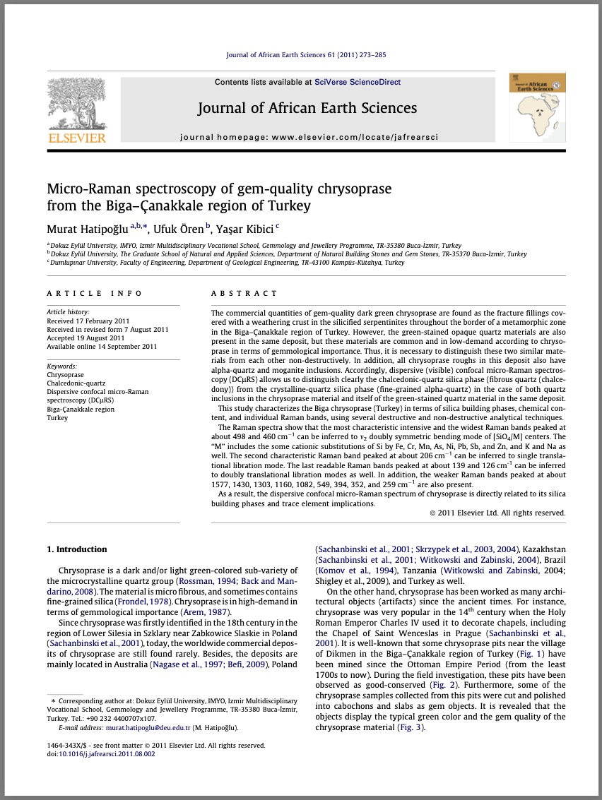
Red gemstones identified mostly as garnets generally occurred in 5th–6th-century Europe as inlay decoration on fine metalwork. The majority are known in cut and mounted form—loose, unmounted pieces are very rarely found. For that reason, it is of great importance that a few years ago seventeen such loose red gemstones were unearthed during a rescue excavation at Hajdúnánás-Fürj-Halom-dűlő (Hajdú-Bihar County). These gemstones are considered remains from a robbed grave in a late 5th-century cemetery, i.e., dated to the Gepidic period. The absence of any related goldsmith’s artifacts and the visible different phases of the gem-cutting process allow us to interpret them as independent pieces in a collection. While mounts and cell walls usually make accurate observation difficult, the loose character of these pieces permitted an extensive investigation to identify the mineral species and determine their possible geological-geographical origin. The results of gemmological-mineralogical and geochemical analyses revealed that the red gemstones are Fe-rich as well as Fe-Mg-rich garnets—i.e., almandine and intermediate varieties between pyrope and almandine. Concerning garnet paragenesis, the raw material experienced medium- to high-grade metamorphism, for the most part at medium pressure, and formed in metapelitic source rocks. The comparative analysis of mineral inclusions and the concentration of major constituents pointed out that they may have been exploited from alluvial deposits most likely in South India and Sri Lanka. An increasing number of archaeometrical investigations are being carried out worldwide to deepen our knowledge about raw material sources and trade links in the Early Middle Ages, when garnet-inlaid jewellery experienced unprecedented spread. Nevertheless, the interpretation of the results presented in this paper can be considered the first provenance study related to early medieval garnets found in present-day Hungary.

Micro-Raman spectroscopy of gem-quality chrysoprase from the Biga-Çanakkale region of Turkey
Journal Article
Hatipoğlu, M., Ören, U. and Kibici, Y. (2011) Micro-Raman spectroscopy of gem-quality chrysoprase from the Biga-Çanakkale region of Turkey. Journal of African Earth Sciences, Vol. 61, pp. 273–285.
Record No. 17219  |  Sinkankas/Born No.: NIS/NIB  |  RWHL
The commercial quantities of gem-quality dark green chrysoprase are found as fracture fillings covered with a weathering crust in the silicified serpentinites throughout the border of a metamorphic zone in the Biga–Çanakkale region of Turkey. However, green-stained opaque quartz materials are also present in the same deposit, but these materials are common and in low demand compared to chrysoprase in terms of gemmological importance. Thus, it is necessary to distinguish these two similar materials from each other non-destructively. In addition, all chrysoprase roughs in this deposit also have alpha-quartz and moganite inclusions. Accordingly, dispersive (visible) confocal micro-Raman spectroscopy (DCμRS) allows us to distinguish clearly the chalcedonic-quartz silica phase (fibrous quartz (chalcedony)) from the crystalline-quartz silica phase (fine-grained alpha-quartz) in the case of both quartz inclusions in the chrysoprase material and the green-stained quartz material in the same deposit. This study characterizes the Biga chrysoprase (Turkey) in terms of silica building phases, chemical content, and individual Raman bands, using several destructive and non-destructive analytical techniques. The Raman spectra show that the most characteristic intensive and the widest Raman bands peaked at about 498 and 460 cm⁻¹ can be inferred to ν₂ doubly symmetric bending modes of [SiO₄/M] centers. The “M” includes some cationic substitutions of Si by Fe, Cr, Mn, As, Ni, Pb, Sb, and Zn, and K and Na as well. The second characteristic Raman band peaked at about 206 cm⁻¹ can be inferred to a single translational libration mode. The last readable Raman bands peaked at about 139 and 126 cm⁻¹ can be inferred to doubly translational libration modes as well. In addition, the weaker Raman bands peaked at about 1577, 1430, 1303, 1160, 1082, 549, 394, 352, and 259 cm⁻¹ are also present. As a result, the dispersive confocal micro-Raman spectrum of chrysoprase is directly related to its silica building phases and trace element implications.

Importance of feather type" inclusions in the identification of natural, treated, synthetic and treated-synthetic yellow sapphire"
Conference Paper
Panjikar, J. and Panjikar, A. (2011) Importance of feather type" inclusions in the identification of natural, treated, synthetic and treated-synthetic yellow sapphire". 32nd International Gemmological Conference, Interlaken, Switzerland pp. 85–88.
Record No. 7768  |  Sinkankas/Born No.: NIS/NIB  |  RWHL

Geochemistry and petrology of nephrite from Alamas, Xinjiang, NW China

Journal Article
Liu, Y., Deng, J., Shi, G., Yui, T.-F., Zhang, G., Abuduwayiti, M., Yang, L. and Sun, X. (2011) Geochemistry and petrology of nephrite from Alamas, Xinjiang, NW China. Journal of Asian Earth Sciences, Vol. 42, No. 3, pp. 440–451.
Record No. 6701  |  DOI: https://doi.org/10.1016/j.jseaes.2011.05.012  |  Sinkankas/Born No.: NIS/NIB  |  RWHL
The Hetian nephrite deposit in Xinjiang, Northwest China, is one of the world’s largest nephrite sources. Within this deposit, the Alamas ore body occurs as veins along the contact between Late Variscan granodiorite and Precambrian dolomitic marble. Petrographic and electron microprobe analyses indicate two potential formation pathways for nephrite: (1) direct transformation of dolomitic marble into tremolite, and (2) dolomitic marble → diopside → tremolite. Nephrite veins display color zoning from white and white-green to green toward the granodiorite contact, corresponding to increasing Cr, FeO, and TFe₂O₃ concentrations. Compared to serpentinite-related nephrite, Alamas nephrite has much lower Cr (8.95–178.7 ppm) and Ni (0.05–3.95 ppm) contents. Bulk-rock REE patterns show strong negative Eu anomalies (0.03–0.21), declining LREE, flat HREE, and low ΣREE (2.84–84.81 ppm), similar to host dolomitic marble (Eu/Eu* = 0.15–0.23, ΣREE = 8.48–11.1 ppm), suggesting a close genetic link. Fluid inclusion homogenization temperatures in tremolite yield a minimum formation temperature of 293 °C. Nephrite δ¹⁸O and δD values range from +3.2 to +6.2‰ and −83.0 to −94.7‰, respectively, with equilibrated fluid δ¹⁸O values of +3.1 to +6.1‰ at 293 °C. These isotopic and petrographic data suggest at least three fluid sources—meteoric water, magmatic water, and CO₂ from dolomite decarbonation—and support a metasomatic, dolomite-related origin for the Alamas ore body.

Analysis of the chromite inclusions found in nephrite minerals obtained from different deposits using SEM-EDS and LRS

Journal Article
Zhang, Z.W. and Gan, F.X. (2011) Analysis of the chromite inclusions found in nephrite minerals obtained from different deposits using SEM-EDS and LRS. Journal of Raman Spectroscopy, Vol. 42, No. 9,, 9/1/11 pp. 1808–1811.
Record No. 6234  |  ISBN/ISSN: 3770486  |  DOI: https://doi.org/10.1002/jrs.2963  |  Sinkankas/Born No.: NIS/NIB  |  RWHL
Scanning electron microscopy with energy dispersive spectrometer (SEM–EDS) and laser Raman spectroscopy (LRS) were used to analyse the chromite inclusions found in four samples of the mineral nephrite that were obtained from Taiwan (one), Manasi in China (two) and New Zealand (one). The chromite inclusions found in sample HL-1 (Taiwan) contain low contents of Mg and Al, as well as a characteristically high Zn content. The most significant finding, however, was that HL-1 could also be distinguished from the other samples by the lower wavenumbers of the positions of the peaks that belong to the A₁g, F₂g(1) and E₉ modes. Compared to the difference in compositions, the difference in the positions of the peaks in the Raman spectra of the chromite inclusions, as revealed by LRS, can more easily be used to distinguish between samples of nephrite that have been obtained from different deposits, and in particular to identify the provenance of the raw material used in ancient nephrite artefacts, in view of the fact that LRS is non-destructive and easy to apply.

Exploring the Literature of Gemstone Inclusions

The literature of gemstone inclusions covers identification, origin determination, treatments, synthesis, mineral inclusions, fluid inclusions and other internal features of natural and synthetic gems.

For a curated selection of important sources, read Inclusions in Gemstones — Hyperion Literature Sources.

About Four Treasures

Four Treasures is Lotus Gemology's freely accessible gemological reference database. Use the full database to search by author, title or keyword and explore literature on gemology, mineralogy, jewelry and related fields. Where available, hyperlinks on titles may lead directly to the full publication.

Search the Complete Four Treasures Database