
Gemstone Inclusions Bibliography

Gemstone Inclusions Bibliography

Explore books, articles and historical sources on inclusions in gemstones in Four Treasures, the Lotus Gemology reference database. This bibliography brings together matching records from the collection, with publication details, abstracts and cover images where available.

Gemstone Inclusion References

The list below matches the terms inclusion or inclusions in the database's searchable fields. Some references discuss inclusions as part of a broader subject.
741–760 of 1,165 matching references

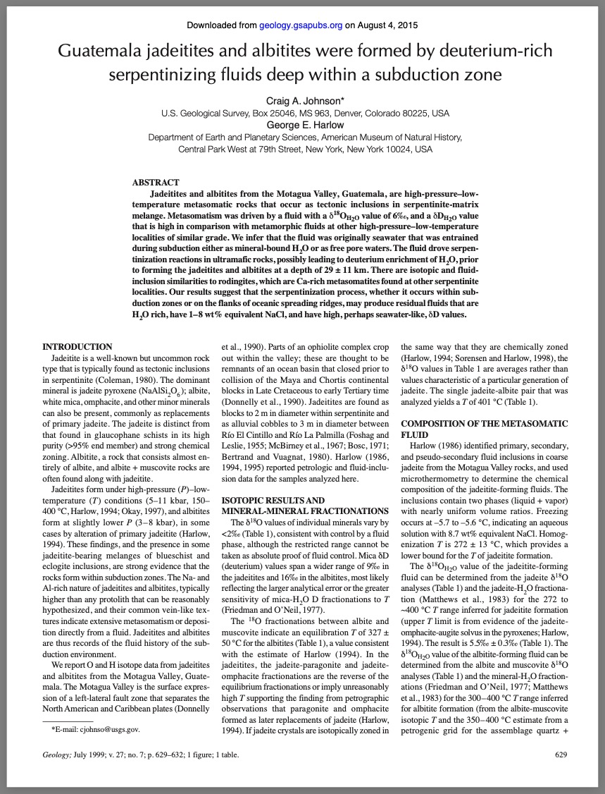
Guatemala jadeitites and albitites were formed by deuterium-rich serpentinizing fluids deep within a subduction-channel
Journal Article
Johnson, C.A. and Harlow, G.E. (1999) Guatemala jadeitites and albitites were formed by deuterium-rich serpentinizing fluids deep within a subduction-channel. Geology, Vol. 27, pp. 629–632.
Record No. 16910  |  DOI: https://doi.org/10.1130/0091-7613(1999)027<0629:GJAAWF>2.3.CO;2  |  Sinkankas/Born No.: NIS/NIB  |  RWHL
Jadeitites and albitites from the Motagua Valley, Guatemala, are high-pressure–low-temperature metasomatic rocks that occur as tectonic inclusions in a serpentinite-matrix mélange. Metasomatism was driven by a fluid with a δ¹⁸Oₕ₂ₒ value of 6‰ and a δDₕ₂ₒ value that is high compared to metamorphic fluids from other high-pressure–low-temperature settings of similar metamorphic grade. It is inferred that the fluid was originally seawater, entrained during subduction either as mineral-bound H₂O or as free pore water. This fluid facilitated serpentinization reactions in ultramafic rocks, potentially leading to deuterium enrichment of the H₂O, before forming the jadeitites and albitites at an estimated depth of 29 ± 11 km. The isotopic signatures and fluid-inclusion data show similarities to rodingites, Ca-rich metasomatic rocks commonly found in serpentinite-bearing environments. These findings suggest that serpentinization, whether in subduction zones or at oceanic spreading ridges, may yield residual fluids that are rich in H₂O, contain 1–8 wt% equivalent NaCl, and display elevated δD values—potentially seawater-like in character.

Corundum from Prilep Dolomitic Marble (Macedonia)
Journal Article
Jeršek, M. and Mirtič, B. (1999) Corundum from Prilep Dolomitic Marble (Macedonia). Scopolia, No. 41, pp. 1–22.
Record No. 7789  |  Sinkankas/Born No.: NIS/NIB  |  RWHL
Structure, chemical composition and solid inclusions exert an influence upon the physical properties of minerals. Besides other parameters they also determine the value of the mineral as a gemstone. In this study we investigated corundum from the dolomitic marble of Prilep (Macedonia), which belongs to the highly metamorphic rocks of the Pelagonian Massif. Marbles show a characteristic mineral paragenesis and include dolomite, calcite, corundum, diaspore, and other accessory minerals. The corundum from Prilep marble appears in a wide number of morphological forms, some of which were identified for the first time in this study. Oriented diaspore intergrowth is also seen in corundum crystals. This affects the physical properties of corundum, such as colour, hardness and density. Diaspore intergrowth can be seen as a silvery glittering in the corundum crystals. We introduce the term “diasporescence” for this phenomenon.

Mineral inclusions in emeralds from different sources
Journal Article
Moroz, I.I. and Eliezri, I.Z. (1999) Mineral inclusions in emeralds from different sources. Journal of Gemmology, Vol. 26, No. 6, pp. 357–363.
Record No. 6047  |  ISBN/ISSN: 221252  |  Sinkankas/Born No.: NIS/NIB  |  RWHL
Mineral inclusions in emeralds from eleven gem-mining regions and a hydrothermally grown synthetic emerald were chemically analysed with the electron microprobe JEOL JXA-8600. Emerald occurrences in Australia, Brazil, Mozambique, Russia, Tanzania (Lake Manyara) and Zambia belong to 'schist type' deposits. The inclusions comprise micas, talc, pyrophyllite, chlorite, wollastonite, chromite, calcite, dolomite, Fe-oxide, apatite, quartz, pyrite, fluorite and plagioclase. The geological settings for Colombian, Nigerian and Sumbawanga (Tanzania) deposits resulted in related assemblages of solid inclusions in these emeralds (for example, chlorine-rich Al- glauconite, sphalerite, titanium-rich mica, anhydrite and beryllium minerals).

Verneuil synthetic corundums with induced “fingerprints”
Journal Article
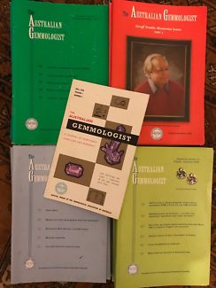
Free, J., Free, I., Brown, G. and T., L. (1999) Verneuil synthetic corundums with induced “fingerprints”. Australian Gemmologist, Vol. 20, No. 8, pp. 342–347.
Record No. 5362  |  Sinkankas/Born No.: NIS/NIB  |  RWHL
Flux-healed, quench-crackled Verneuil synthetic ruby and sapphire are being produced in Chanthaburi, Thailand. First, the Verneuil boules are heated and then plunged into water to generate the quench-crackled effect. After the boules have been cobbed to yield small facetable fragments, these pieces are heat treated in an unspecified colorless flux for two days in a kerosene-fired kiln. The process is completed with a one-day heat treatment in an acetylene-fired kiln. The key identifying features are curved color banding or striae, a “checkerboard” pattern of flux-filled fractures, and increased transparency to shortwave UV radiation.

Ruby from Tunduru-Songea, East Africa: Some basic observations
Journal Article
Hamid, G., Kelly, S.M.B. and Brown, G. (1999) Ruby from Tunduru-Songea, East Africa: Some basic observations. Australian Gemmologist, Vol. 20, No. 8, pp. 326–330.
Record No. 5361  |  Sinkankas/Born No.: NIS/NIB  |  RWHL
Gem corundum with a bright pink to purplish red hue is recovered from river gravels in the Tunduru–Songea area of southern Tanzania, and sold in Bangkok. Most, but not all, of these rubies appear to have been heat treated prior to sale. The rubies have refractive indices of nε = 1.760–1.765 and nω = 1.768–1.773 (yielding a birefringence of 0.008–0.009) and specific gravity values of 3.95–4.04. The characteristic inclusions found in both the obviously heat-treated and minimally heat-treated stones are described.

Texture formation and element partitioning in trapiche ruby
Journal Article
Sunagawa, I., Bernhardt, H.-J. and Schmetzer, K. (1999) Texture formation and element partitioning in trapiche ruby. Journal of Crystal Growth, Vol. 206, pp. 322–330.
Record No. 5359  |  Sinkankas/Born No.: NIS/NIB  |  RWHL
Trapiche rubies have six red growth sectors separated by six opaque yellow to white dendritic (i.e., branching) “arms” that run from the central core to the six corners of the hexagonal crystal (see Summer 2000 Gem News, pp. 168–169). This article explores the trace-element chemistry and origin of these unusual textural features, which are found in some specimens from Mong Hsu, Myanmar.. X-ray microfluorescence and electron microprobe analyses, as well as microscopic examinations, on polished slabs cut perpendicular to the c-axis showed both chemical and growth zoning. The opaque arms consist of ruby with heterogeneous concentrations of solid (e.g., calcite and dolomite), liquid, or two-phase inclusions. Zoning of trace elements (particularly chromium) revealed differential rates of crystal growth. The authors conclude that the opaque arms formed first during a period of rapid growth; the red growth sectors formed during a subsequent period of slower, layer-by-layer growth. The rapid growth implied by trace-element partitioning in the arms was controlled by temperature. The growth of the interstitial ruby phases was governed by the kinetic factors dictated by the dendritic branches.JL

Les rubis d’Ishtar: Étude archéologique
The rubies of Ishtar: Archaeological study
Book Section
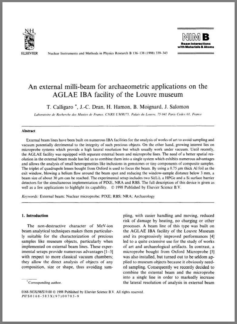
Tallon, F. (1999) Les rubis d’Ishtar: Étude archéologique. [The rubies of Ishtar: Archaeological study] In: Cornaline et pierres precieuses, La Mediterranee de l'Antiquité a l’Islam, Paris: Louvre éditions, pp. 229–234.
Record No. 3297  |  ISBN/ISSN: 9782110000000  |  Sinkankas/Born No.: NIS/NIB  |  Language: in French  |  RWHL
For their aesthetic and symbolic value, gemstones have always been sought out and used by humans. The Mesopotamian tablets, the oldest written records, frequently mention gemstones: lapis lazuli, carnelian… However, due to their rarity, the hard precious stones of Antiquity (diamond, ruby, emerald, sapphire) have been less studied. The examination of the Parthian statuette preserved in the Louvre Museum, an object inlaid with rubies, provided an opportunity for the Laboratory of Research of the Museums of France (LRMF) to implement ion beam analysis methods for in-depth research in the field of historical gemmology. The archaeological piece at the core of this study was discovered in Mesopotamia. Based on typology, it is attributed to the Parthian period and would thus date to the 3rd century BCE. This remarkable object represents the goddess Ishtar, associated with Aphrodite. The statuette is made of alabaster; her eyes and navel are inlaid with a red stone initially thought to be glass (fig. 1). In 1995, during the dossier-exhibition Precious Stones of the Ancient East, from the Sumerians to the Sasanians organized by François Talion at the Louvre Museum, Annie Caubet, curator in charge of the Department of Near Eastern Antiquities, proposed that we study this statuette. The objectives were, on the one hand, to confirm the suggestion of S. Van Roy², who believed these cabochons to be rubies, and on the other hand, to attempt to determine the geographic origin of these gems. The study of the rubies on the Parthian statuette from the Louvre Museum combines optical observation of their inclusions with their geochemical characterization using the PIXE method (with the help of AGLAE). It first confirmed that the red inlays of the eyes and navel are indeed natural rubies. Furthermore, it provided an opportunity to build a compositional database for major ruby deposits. Finally, thanks to statistical analysis, we can propose Burma as the most probable origin of the statuette’s rubies, though Sri Lanka cannot be ruled out. These results, along with recent improvements to the AGLAE accelerator, pave the way for a complementary study to strengthen the attribution and attempt to decide between a Burmese or Sri Lankan origin for these rubies. Indeed, the new extracted beam from AGLAE now makes it possible to focus the beam on an area just 0.03 mm in diameter. This tool is well suited for characterizing the mineral inclusions observed in our study (rutile, zircons, phlogopite…), a key factor for authenticating the paragenesis and, consequently, for tracing the provenance of the gems. These gemstones are evidence of exchange or trade across several thousand kilometers between Mesopotamia and Southeast Asia in the 3rd century BCE. To our knowledge, this is the earliest known use of ruby in this region. This representation of Ishtar is remarkable because it combines influences from three civilizations: Mesopotamia through the deity represented, Greece through its Hellenistic style, and Asia through both the placement (eyes and navel) and the nature of the precious stones.

An external milli-beam for archaeometric applications on the AGLAE IBA facility of the Louvre museum
Journal Article
Calligaro, T., Dran, J.C., Hamon, H., Moignard, B. and Salomon, J. (1998) An external milli-beam for archaeometric applications on the AGLAE IBA facility of the Louvre museum. Nuclear Instruments and Methods in Physics Research B, Vol. B138, pp. 339–343.
Record No. 27058  |  DOI: https://doi.org/10.1016/S0168-583X(97)00703-9  |  Sinkankas/Born No.: NIS/NIB  |  RWHL
External beam lines have been built on numerous IBA facilities for the analysis of works of art to avoid sampling and vacuum potentially detrimental to the integrity of such precious objects. On the other hand, growing interest lies on microprobe systems which provide a high lateral resolution but which usually work under vacuum. Until recently, the AGLAE facility was equipped with separate external beam and microprobe lines. The need of a better spatial resolution in the external beam mode has led us to combine them into a single system which exhibits numerous advantages and allows the analysis of small heterogeneities like inclusions in gemstones or tiny components of composite samples. The triplet of quadrupole lenses bought from Oxford is used to focus the beam. By using a 0.75 μm thick Al foil as the exit window, blowing a helium flow around the beam spot and reducing the window–sample distance below 3 mm, a beam size of about 30 μm can be reached. The experimental setup includes two Si(Li), a HPGe, and a Si surface barrier detector for the simultaneous implementation of PIXE, NRA, and RBS. The full description of this device is given as well as a few applications to highlight its capability.

The chemical signature of emeralds from the Campos Verdes-Santa Terezinha Mining District, Goiás, Brazil
Journal Article
Pulz, G.M., Silva, L.J.H.d.e.-R., Neto, L.S.B., Brum, T.M.M., Juchem, P.L., Santos, C.A., Pereira, V.P. and Silva, J.J. (1998) The chemical signature of emeralds from the Campos Verdes-Santa Terezinha Mining District, Goiás, Brazil. Journal of Gemmology, Vol. 26, No. 4, October, pp. 252–261.
Record No. 6063  |  ISBN/ISSN: 221252  |  Sinkankas/Born No.: NIS/NIB  |  RWHL
Emerald crystals from the Campos Verdes mining district have been analysed by inductively coupled plasma mass spectrometry (ICP-MS), Fourier transform infrared spectroscopy and Mössbauer 2+ 3+ spectroscopy. These gems contain measurable amounts of Fe , Fe , Cr, V, Sc, Ni and Zn; large-ion lithophile element (LILE, Ba, Sr, Rb, Pb) and high field strength element (HFSE, Zr, Y, Ti) contents are near or below the limit of detection. This chemical signature reflects the metasomatic reactions between the mineralizing fluids and the ultrabasic host-rocks of this gem deposit.

Lab Notes: Jadeite with inclusions of zircon
Journal Article
Koivula, J.I. and Moses, T. (1998) Lab Notes: Jadeite with inclusions of zircon. Gems & Gemology, Vol. 34, No. 1, Spring, p. 45.
Record No. 5496  |  ISBN/ISSN: 0016626X  |  Sinkankas/Born No.: NIS/NIB  |  RWHL
As the practice of bleaching and impregnating jadeite has become more common over the last few years, the amount of jadeite we test in the laboratory has increased signifi-cantly. During the microscopic exam-ination of one variegated green jadeite cabochon for possible evidence of treatment, we noted that the stone contained a number of small crystal inclusions, some of which reached the surface (figure 4). These surface-reaching crystals appeared to be somewhat harder than the surround-ing jadeite, and in reflected light they had a higher surface luster. With the recent acquisition of GIA’s Renishaw Raman Imaging Microscope System, we were able to examine two of these crystals nondestructively. By focusing the argon laser through the 50 x objective of the Leica targeting microscope and run-ning several continuous scans, we obtained clear spectra of the two “inclusions.” A computer search of our data base revealed that these spec-tra matched the spectrum for zircon. Visible characteristics of the inclu-sions (e.g., crystal form, color, trans-parency, and relative hardness) were consistent with this identification. This is the first time that we have encountered zircon crystals as inclu-sions in jadeite. Coincidentally, about a month prior to our discovery, a zir-con inclusion in a jadeite cabochon was also identified by means of the same technique at the Center for Gemstone Testing in Bangkok (Ken-neth Scarratt, pers. comm.).

Models of corundum origin from alkali basaltic terrains: a reappraisal
Journal Article
Sutherland, L.F., Hoskin, P.W.O., Fanning, C.M. and Coenraads, R.R. (1998) Models of corundum origin from alkali basaltic terrains: a reappraisal. Contributions to Mineralogy and Petrology, Vol. 133, No. 4, pp. 356–372.
Record No. 5294  |  ISBN/ISSN: 107999  |  DOI: https://dx.doi.org/10.1007/s004100050458  |  Sinkankas/Born No.: NIS/NIB  |  RWHL
Corundums from basalt fields, particularly in Australia and Asia, include a dominant blue-green-yellow zoned magmatic suite (BGY suite) and subsidiary vari-coloured metamorphic suites. The BGY corundums have distinctive trace element signatures, with up to 0.04 wt% Ga₂O₃ and low Cr/Ga and Ti/Ga ratios (<1). Different melt origins for the BGY suite are examined based on inclusion and intergrowth mineralogy, petrologic associations, and tectonic setting. Over 100 analysed primary mineral inclusions include typical feldspars, zircon, Nb–Ta oxides, hercynite–magnetite, gahnospinel, rutile–ilmenite solid solution, calcic plagioclase, Ni-rich pyrrhotite, thorite, and low-Si, Fe-rich glassy inclusions. This expands upon prior surveys; New England, eastern Australia, in particular, shows the most diverse inclusion suite yet documented from basalt-hosted corundums (20 phases). Rare earth element patterns of zircon inclusions, intergrowths, and megacrysts are similar, with the exception of variable Eu depletion. Temperature estimates derived from magnetite exsolution textures, feldspar compositions, and fluid inclusion homogenization suggest corundum crystallization occurred at 685–900 °C. The overlap in Nb–Ta oxide compositions between corundum inclusions and those found in niobium–yttrium–fluorine (NYF) granitic pegmatites supports a silicate melt origin. The MELTS thermodynamic model was applied to amphibole–pyroxenite xenolith compositions from basalts to test the viability of corundum formation from mantle-derived melts. The calculations indicate corundum crystallization occurs at 720–880 °C and 0.7–1.1 GPa, producing residual feldspathic assemblages matching observed mineral inclusions and xenoliths. A model is thus proposed in which corundum in the BGY suite forms from low-volume silicate melts generated during partial melting of amphibole-bearing lithospheric mantle under the influence of magmatic plume activity.

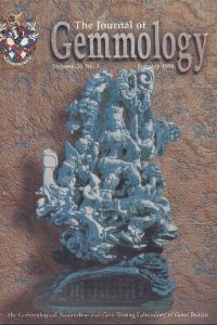
An interdisciplinary approach to identifying solid inclusions in corundum: thorite in a ‘Sri Lanka’ sapphire
Journal Article
Carbonin, S., Sbrignadello, G. and Ajo, D. (1998) An interdisciplinary approach to identifying solid inclusions in corundum: thorite in a ‘Sri Lanka’ sapphire. Journal of Gemmology, Vol. 26, No. 4, October, pp. 262–264.
Record No. 4732  |  ISBN/ISSN: 221252  |  Sinkankas/Born No.: NIS/NIB  |  RWHL

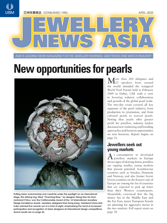
Spinel and garnet star networks: An interesting asterism in gems from Sri Lanka
Journal Article
Kumaratilake, W.L.D.R.A. (1998) Spinel and garnet star networks: An interesting asterism in gems from Sri Lanka. Journal of Gemmology, Vol. 26, No. 1, January, pp. 24–28.
Record No. 4144  |  ISBN/ISSN: 221252  |  Sinkankas/Born No.: NIS/NIB  |  RWHL
Four-ray and six-ray star networks are reported in spinel and garnet from Sri Lanka. Details of their arrangement and orientations are discussed.

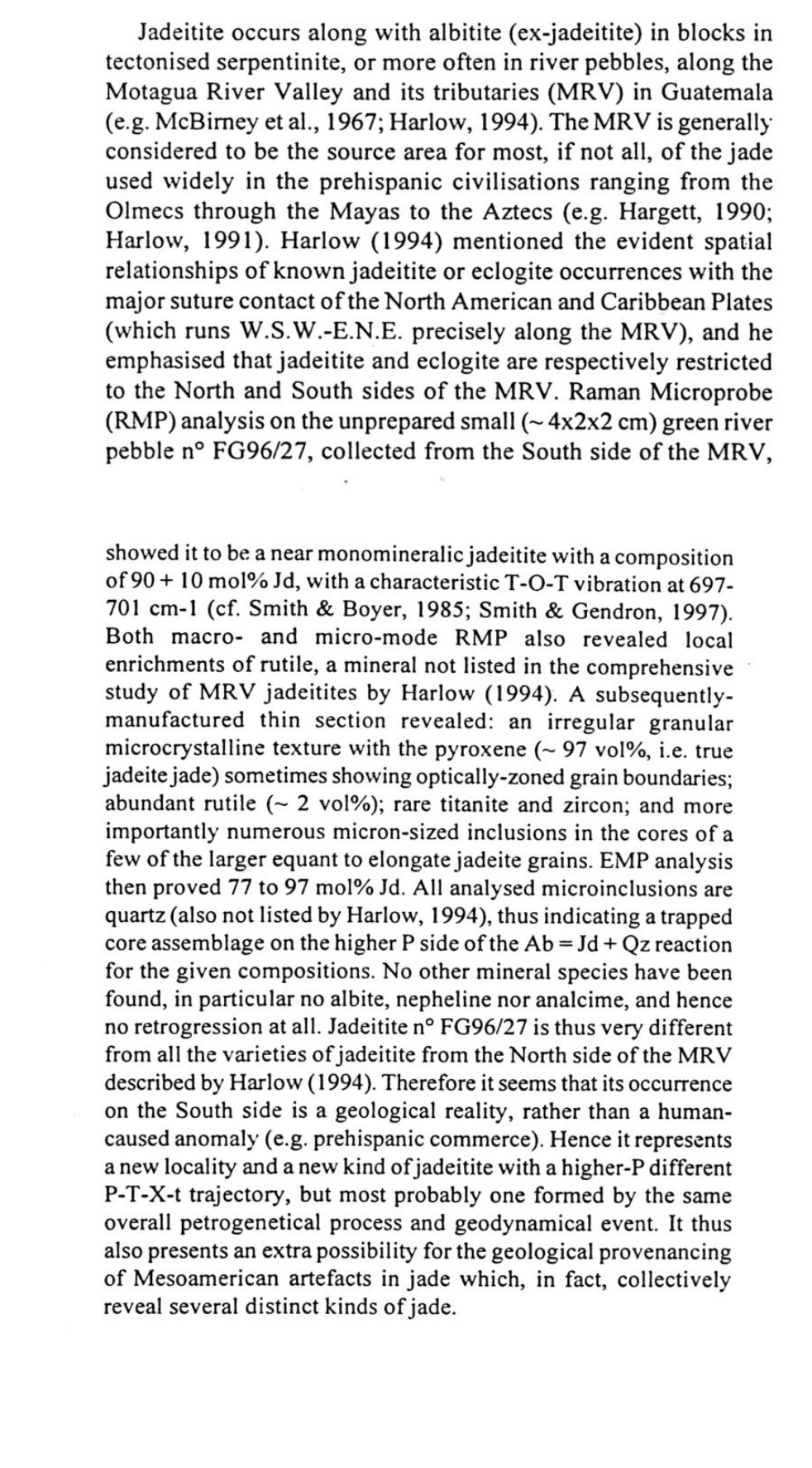
China company produces hydrothermal rubies
Journal Article
Anonymous (1998) China company produces hydrothermal rubies. Jewellery News Asia, No. 169, p. 50.
Record No. 2950  |  Sinkankas/Born No.: NIS/NIB  |  RWHL
Hydrothermal synthetic rubies marketed under the name “JL synthetic rubies” are available from Guangxi Jewelry Co. Ltd. in Guilin, China (Guangxi Province). The material ranges from red to orange-red in color, with some displaying purple hues. The synthetic rubies fluoresce strong red to long-wave ultraviolet radiation; the fluorescence is less intense to short-wave UV. Colorless “net-like” inclusions are reported. The synthetic rubies have R.I. values of 1.759–1.769, and an S.G. of 4.01. They are incorporated into gold rings, pendants, earrings, and bracelets. MM

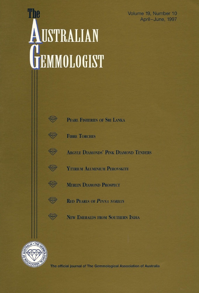
New Locality and a New Kind of Jadeite Jade from Guatemala: Rutile-Quartz-Jadeite
Conference Paper
Smith, D.C. and Gendron, F. (1997) New Locality and a New Kind of Jadeite Jade from Guatemala: Rutile-Quartz-Jadeite. Fifth International Eclogite Conference, Terra Nova 9, p. 35.
Record No. 28500  |  Sinkankas/Born No.: NIS/NIB  |  RWHL
Jadeitite occurs along with albitite (ex-jadeitite) in blocks in tectonised serpentinite, or more often in river pebbles, along the Motagua River Valley and its tributaries (MRV) in Guatemala (e.g. McBirney et al., 1967; Harlow, 1994). The MRV is generally considered to be the source area for most, if not all, of the jade used widely in the prehispanic civilisations ranging from the Olmecs through the Mayas to the Aztecs (e.g. Hargett, 1990; Harlow, 1991). Harlow (1994) mentioned the evident spatial relationships of known jadeitite or eclogite occurrences with the major suture contact of the North American and Caribbean Plates (which runs W.S.W.-E.N.E. precisely along the MRV), and he emphasised that jadeitite and eclogite are respectively restricted to the North and South sides of the MRV. Raman Microprobe (RMP) analysis on the unprepared small (~4×2×2 cm) green river pebble n° FG96/27, collected from the South side of the MRV, showed it to be a near monomineralic jadeitite with a composition of 90 ± 10 mol% Jd, with a characteristic T-O-T vibration at 697–701 cm⁻¹ (cf. Smith & Boyer, 1985; Smith & Gendron, 1997). Both macro- and micro-mode RMP also revealed local enrichments of rutile, a mineral not listed in the comprehensive study of MRV jadeitites by Harlow (1994). A subsequently manufactured thin section revealed: an irregular granular microcrystalline texture with the pyroxene (~97 vol%, i.e. true jadeite jade) sometimes showing optically-zoned grain boundaries; abundant rutile (~2 vol%); rare titanite and zircon; and more importantly numerous micron-sized inclusions in the cores of a few of the larger equant to elongate jadeite grains. EMP analysis then proved 77 to 97 mol% Jd. All analysed microinclusions are quartz (also not listed by Harlow, 1994), thus indicating a trapped core assemblage on the higher P side of the Ab = Jd + Qz reaction for the given compositions. No other mineral species have been found, in particular no albite, nepheline nor analcime, and hence no retrogression at all. Jadeitite n° FG96/27 is thus very different from all the varieties of jadeitite from the North side of the MRV described by Harlow (1994). Therefore it seems that its occurrence on the South side is a geological reality, rather than a human-caused anomaly (e.g. prehispanic commerce). Hence it represents a new locality and a new kind of jadeitite with a higher-P different P-T-X-t trajectory, but most probably one formed by the same overall petrogenetical process and geodynamical event. It thus also presents an extra possibility for the geological provenancing of Mesoamerican artefacts in jade which, in fact, collectively reveal several distinct kinds of jade.

New emerald deposits from Southern India
Journal Article
Panjikar, J., Ramchandran, K.T. and Balu, K. (1997) New emerald deposits from Southern India. Australian Gemmologist, Vol. 19, No. 10, April–June, pp. 427–432.
Record No. 26923  |  Sinkankas/Born No.: NIS/NIB  |  RWHL
In the mid-1990s, emeralds were discovered embedded in the wall of a well at the village of Sankari Taluka, in southern India’s state of Tamil Nadu. Gemmological research into the chemistry, properties, and characteristic inclusions of these emeralds has revealed similarities between the properties of Sankari emerald and those of emerald from several previously described locations on the island of Madagascar. This finding suggests that Sankari emeralds may have been emplaced into the ancient supercontinent of Gondwanaland before India rifted from it some 200 million years ago.

Gem News: Sapphires from northern Madagascar
Journal Article
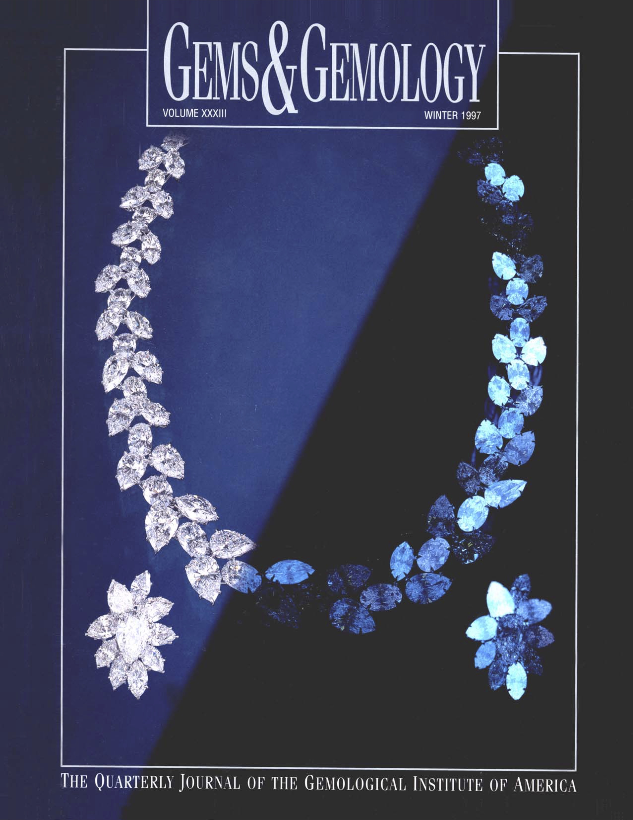
Johnson, M.L. and Koivula, J.I. (1997) Gem News: Sapphires from northern Madagascar. Gems & Gemology, Vol. 33, No. 4, Winter, p. 305.
Record No. 6005  |  ISBN/ISSN: 0016626X  |  Sinkankas/Born No.: NIS/NIB  |  RWHL
Sapphires from northern Madagascar. In the past few years, several new and important sapphire deposits have been discovered in Madagascar. Dr. Margherita Superchi, of CISGEM, Milan, Italy, described a new occurrence at Ambondromifehy, in Antsiranana Province, in the northern part of the island. The sapphires found to date were alluvial, having been weathered from basaltic rocks that were emplaced intermittently during the period from the Upper Oligocene to the Pleistocene. The sapphires are thought to be xenocryts; that is, they did not crystallize from the basaltic magma, but simply were transported by it. They are associated with zircon, spinel, pyroxene, and olivine. Dr. Superchi and her colleagues (A. Donini, D. Muzzioli, and E. Roman) obtained detailed gemological data, chemical compositions, and optical and Raman spectra for 74 crystals (the largest being 182.59 ct) and 13 cut stones. Among the more interesting discoveries were inclusions of pyrochlore, zircon, and sodic plagioclase, which, with the chemical data (e.g., high gallium contents) confirm the “basaltic” association of these sapphires (see the earlier abstract on gem corundum).

Phase transformation of TiO₂ precipitates in sapphire (α-Al₂O₃) induced by the loss of coherency
Journal Article
Xiao, S.Q., Dahmen, U. and Heuer, A.H. (1997) Phase transformation of TiO₂ precipitates in sapphire (α-Al₂O₃) induced by the loss of coherency. Philosophical Magazine A, Vol. 75, No. 1, pp. 221–238.
Record No. 5529  |  Sinkankas/Born No.: NIS/NIB  |  RWHL
The first phase to precipitate in Ti-doped sapphire (Ti:α-Al₂O₃) single crystals annealed at 1300 °C in air is a high-pressure form of TiO₂ with the α-PbO₂ structure (α-TiO₂). The coherent precipitates form as oblate spheroidal plates with habit planes parallel to (0001). Because of a 5% lattice mismatch with sapphire, the small coherent α-TiO₂ precipitates are under an effective compressive stress, which stabilizes the high-pressure polymorph. During coarsening, the particles lose coherency by emitting a dislocation loop with a f(1̄101) Burgers vector, which reduces the stress on the particles and allows them to transform to the rutile structure. The orientation relationship between the sapphire matrix, α-TiO₂ and rutile has been determined and a lattice correspondence for the phase transformations established. Although the transformation from α-TiO₂ to rutile is purely structural, its rate is controlled by atomic diffusion; the increased atomic volume of rutile compared with α-TiO₂ is accommodated by expelling extra mass of Al₂O₃ into nearby interstitial dislocation loops and is associated with a change in the precipitate morphology.

A Raman microscope in the gemmological laboratory: First experiences of application
Journal Article
Hänni, H.A., Kiefert, L. and Chalain, J.P. (1997) A Raman microscope in the gemmological laboratory: First experiences of application. Journal of Gemmology, Vol. 25, No. 6, April, pp. 394–406.
Record No. 5371  |  ISBN/ISSN: 221252  |  Sinkankas/Born No.: NIS/NIB  |  RWHL
CMS: The Swiss Gemmological Institute (SSEF) in Basel has found its recent acquisition of a Raman microscope well worth the investment, to judge by this excellent account of its initial applications at this laboratory. Much more accurate, easier to use, and considerably less expensive than its predecessors, the Renishaw Raman microscope at SSEF easily fits on a desktop. It has enabled SSEF staff members to identify CO₂ inclusions in sapphire that are diagnostic of heat treatment, detect and identify fracture fillings in emerald, easily identify mounted gemstones, and characterize specimens with irregular surfaces. It can also distinguish component minerals in many aggregates. The method is nondestructive in all but a few well-documented conditions and works on all types of materials, except those having simple compositions and high symmetry, such as metals and alloys. Raman spectroscopy does require that the user be aware of its limitations and parameters; also, the lack of a large, published database means that each laboratory must obtain many of its own reference spectra from known samples. However, Raman spectroscopy clearly has earned a place of respect among the few high-tech laboratory techniques that have proved indispensable to gemological testing today.

Micro-PIXE analysis of trace element concentrations of natural rubies from different locations in Myanmar

Journal Article
Sanchez, J.L., Osipowicz, T., Tang, S.M., Tay, T.S. and Win, T.T. (1997) Micro-PIXE analysis of trace element concentrations of natural rubies from different locations in Myanmar. Nuclear Instruments and Methods in Physics Research B, Vol. 130, pp. 682–686.
Record No. 5368  |  Sinkankas/Born No.: NIS/NIB  |  RWHL
The authors analyzed 130 untreated rough rubies from nine metamorphic localities in Myanmar, including Mogok and Mong Hsu, using micro proton-induced X-ray emission (PIXE). The concentrations of chromium, titanium, vanadium, iron, copper, and gallium—the most abundant trace elements—were determined for a 200 × 200 μm area on each stone. In a unique approach, the authors used the trace-element mapping capability of the instrument to eliminate inhomogeneities, such as inclusions, from the analyses for the areas examined. The measurements revealed that, with the exception of gallium, there were large variations in the trace-element chemistry of rubies from the different localities, as well as for rubies from the same locality. Nevertheless, some consistent differences were also found. Using statistical analysis, including tree clustering and factor analysis, the authors determined that the origin of rubies from six of the nine localities could, indeed, be determined using this technique.

Exploring the Literature of Gemstone Inclusions

The literature of gemstone inclusions covers identification, origin determination, treatments, synthesis, mineral inclusions, fluid inclusions and other internal features of natural and synthetic gems.

For a curated selection of important sources, read Inclusions in Gemstones — Hyperion Literature Sources.

About Four Treasures

Four Treasures is Lotus Gemology's freely accessible gemological reference database. Use the full database to search by author, title or keyword and explore literature on gemology, mineralogy, jewelry and related fields. Where available, hyperlinks on titles may lead directly to the full publication.

Search the Complete Four Treasures Database